
Presentation
Right knee pain.
Patient Data

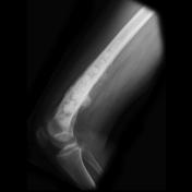

The distal half of the femur is occupied and expanded with a heterogeneous mass with areas of bone formation. Posterior the periosteum is elevated (Codman's triangle).
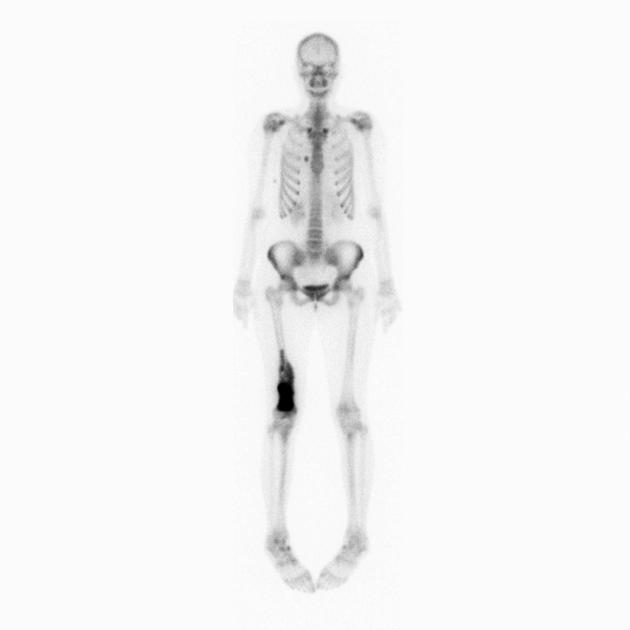
Delayed phase bone scan image demonstrates intense uptake in the distal right femur. A single focus of increased attenuation projects just to the right of the sternum inline with the chondral cartilage of the third rib. No other suspicious lesions identified.

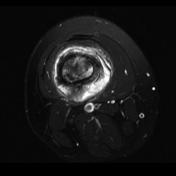
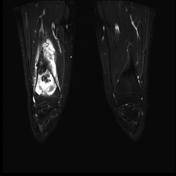
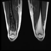
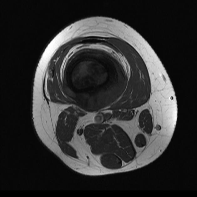
MRI through the lesion demonstrates a soft tissue mass completely replacing the medullary cavity and extending into the adjacent soft tissues. It is low to intermediate in signal on T1 and high on T2 weighted images.
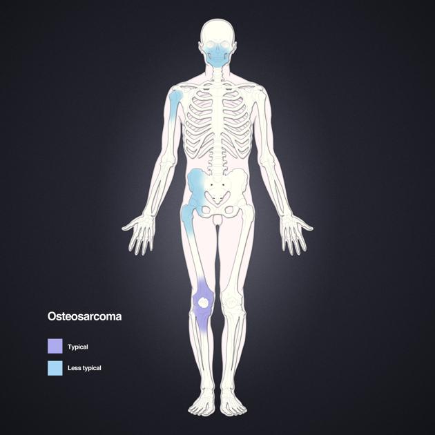
Distribution of osteosarcomas. The most frequent location is around the knee. Layout and distribution: Frank Gaillard 2009, Line drawing of skeleton: Patrick Lynch 2006, Creative Common NC-SA-BY
This patient went on to have surgery and an osteosarcoma was histologically confirmed.
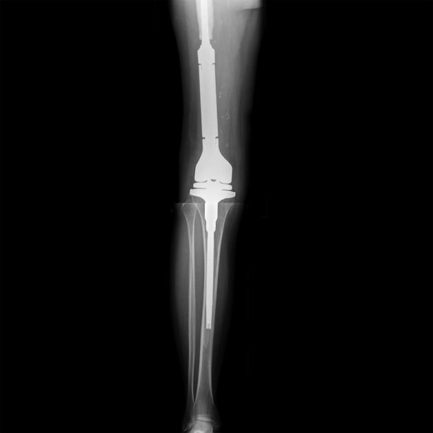
A limb sparing resection has been performed. The distal femur and adjacent tibial plateau has been resected and a total knee replacement inserted.
Case Discussion
This case demonstrates relatively typical appearances of a bone forming osteosarcoma of the distal femur (typical location) in a 17-year-old (typical age).


 Unable to process the form. Check for errors and try again.