
Presentation
Primary infertility.
Patient Data
Age: 40 years
Gender: Female
From the case:
Ovarian fibroma - uterine leiomyomas


Download
Info
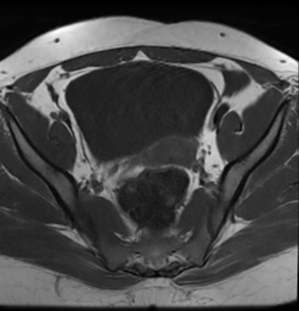
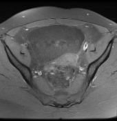
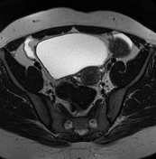
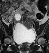
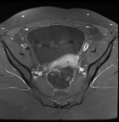
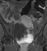
There is a well-defined left ovarian mass measuring (6.2 X 5.8 X 4.8 cm) of isosignal to the myometrium on T1, low signal on T2 with central areas of low signal on T1 and high signal on T2 (areas of edema or cystic degeneration). The postcontrast sequences show a heterogeneous enhancement.
Small simple unilocular cyst of the right ovary (3 cm).
Two small intramural leiomyomas are noted.
Mild peritoneal effusion.
Case Discussion
MRI features of a unilateral ovarian mass of T2 low signal with central areas of edema / cystic degeneration and heterogeneous enhancement in middle-aged women suggest probably an ovarian fibroma.
On imaging, the main differential diagnosis is thecoma and fibrothecoma which:
- most occur in adult women, with ~66% in postmenopausal women
- tend to have a brighter signal on T2 given edema and cystic degeneration
- contrast-enhancement may be observed given the vascularization of the theca cells


 Unable to process the form. Check for errors and try again.