
Presentation
Abdominopelvic pain and progressive distension.
Patient Data
Age: 40 years
Gender: Female
From the case:
Ovarian serous cystadenocarcinoma


Download
Info
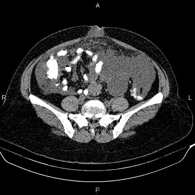
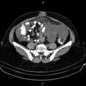
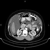
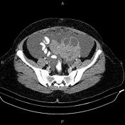
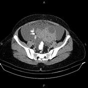
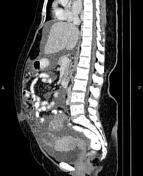
There is a 150 × 105 × 105 mm cystic and solid mass in the left adnexa. There is also a 50 × 40 × 45 mm multiseptated cystic lesion at right adnexa. The ovaries couldn’t be defined separate from these lesions.
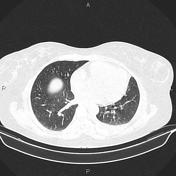
Massive ascites is evident accompanied by omental thickening. Additionally, a small pleural effusion is present on the left side.
Case Discussion
Adnexal masses; pathology proven ovarian serous cystadenocarcinoma with massive ascites and omental thickening.
Ovarian serous cystadenocarcinoma is the malignant form of ovarian serous tumor, the most common type of ovarian epithelial tumor. It is the most common type of ovarian malignancy.


 Unable to process the form. Check for errors and try again.