
Presentation
Pelvic pain and palpable mass on physical exam.
Patient Data
Age: 45 years
Gender: Female
From the case:
Ovarian serous cystadenocarcinoma


Download
Info
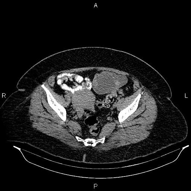
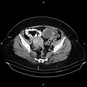
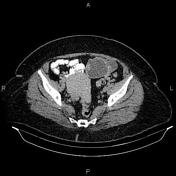
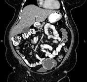
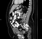
A 93×58 mm cystic lesion with thick wall and septations, foci of calcifications and enhancing solid components is seen at left adnexa. The left ovary couldn’t be defined separate than mentioned cystic lesion. There is no sign of local invasion to adjacent structures and no regional lymphadenopathies.
The uterus contains a few small fibroids.
Several non-enhanced simple cortical cysts are seen at both kidneys.
Case Discussion
Left adnexal complex cystic mass; pathology proven ovarian serous cystadenocarcinoma which is the malignant form of ovarian serous tumor, the most common type of ovarian epithelial tumor. It is the most common type of ovarian malignancy.


 Unable to process the form. Check for errors and try again.