
Presentation
severe pelvic pain of 3 days duration. History of right ovariectomy for ovarian cyst (surgical report not available).
Patient Data
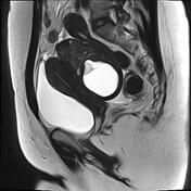

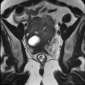

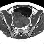
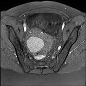
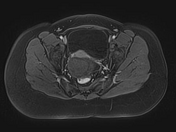

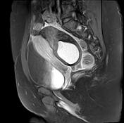

Large well-circumscribed ovoid retrouterine mass located in Douglas pouch. It elicits an intermediate signal on T1 and T2 with numerous peripheral tiny cysts (follicles) of high T2 signal as well as a cystic area of high signal on T1 and T2 (hemorrhage). No enhancement on postcontrast dynamic sequences as well as on delayed sequences. The twisted pedicle well-demonstrates on T2 sequences.
Minimal effusion is seen in the Douglas pouch.
The left ovary is not visualized (history of ovariectomy).
Case Discussion
The clinical presentation and the MRI features are most consistent with an ovarian torsion that was confirmed at the surgery.


 Unable to process the form. Check for errors and try again.