
Presentation
Right-sided neck swelling for 4 -6 weeks. No pain or fever.
Patient Data
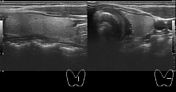


The right lobe of the thyroid gland is replaced by a large heterogeneous echopattern solid lesion. The lesion is isoechoic as well as hypoechoic area without cystic area. There are multiple tiny echogenic foci in the lesion - tiny calcifications. There is no hypoechoic halo around the lesion. There is no capsular breach. Size of the lesion is 58 x 35 x 27 mm. It is wider than taller. There are significant flow signals in the lesion. Isthmus and the left lobe of the thyroid gland shown normal size and echopattern without any focal lesion.
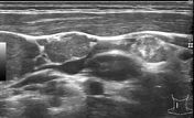
There are right level III-IV enlarged, non-necrotic nodes with tiny calcific foci. The nodes show increased vascularity. There is no periadenitis.
Case Discussion
An adult female presented with right-sided cervical lumps for the last few weeks. Neck ultrasound revealed right lobe thyroid gland lesion with ultrasound findings favoring papillary thyroid cancer. There was right-sided cervical lymphadenopathy with tiny calcification favoring metastatic spread from thyroid lesion.
FNAC from thyroid gland suggested papillary thyroid carcinoma.


 Unable to process the form. Check for errors and try again.