
Presentation
Patient with a very long history of exhaustion and easily fatigability.
Patient Data

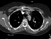

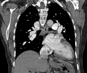
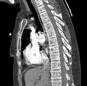
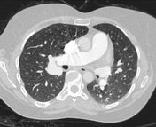
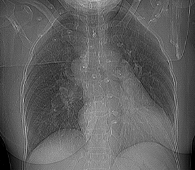
A scout view shows an abnormal rounded radiopaque shadow in the right hilar region to the right of the distal trachea. The CT scan reveals a large anomalous venous trunk that ended in the SVC and drained most of the right lung, a feature consistent with partial anomalous pulmonary venous return.
The right inferior pulmonary vein drains some segment of the right lung and normally ends in the left atrium. The left pulmonary vein also drains generally to the left atrium via a common short trunk.
Dilated right cardiac chambers, pulmonary artery and its branches.
Case Discussion
A case of Supracardiac type partial anomalous pulmonary venous return with right heart volume overload. A small patent foramen ovale was noticed in the echocardiography (not shown).


 Unable to process the form. Check for errors and try again.