

Illustrations of common bone bruise patterns, associated soft tissue injuries and MRI appearance including: 1) lateral patellar dislocation, 2) dashboard injury, 3) hyperextension, 4) valgus force, and 5) pivot shift.
Author: Stefan Tigges.
Case Discussion
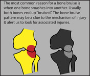
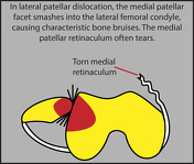
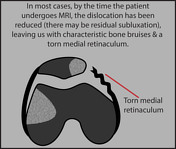
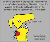
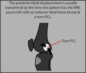
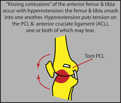
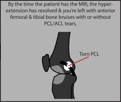
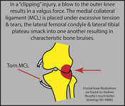
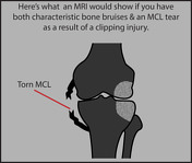
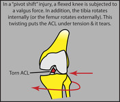
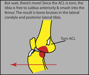
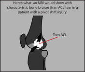
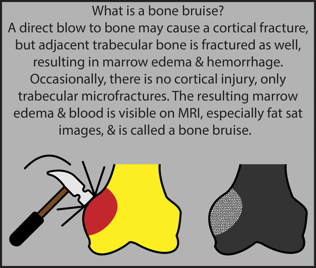
Bone bruises are trabecular microfractures with accompanying hemorrhage that occur when bone is violently compressed and are frequently seen in a variety of knee injuries. The pattern of bone bruises may indicate the mechanism of injury and can be used to predict concomitant soft tissue abnormalities. Five different bone bruise patterns have been described:
lateral patellar dislocation
dashboard injury
hyperextension
valgus force
pivot shift
These diagrams walk you through how these injuries occur, the associated soft tissue injuries, and the appearance on fat suppressed MR images. Remember that not all bone bruises will conform to these patterns, and even patients with characteristic bone bruise patterns may not have the expected soft tissue injuries.


 Unable to process the form. Check for errors and try again.