Presentation
Acute onset of left lower quadrant abdominal pain with palpable left iliac fossa mass.
Patient Data
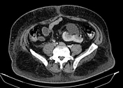

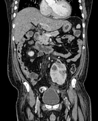

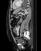

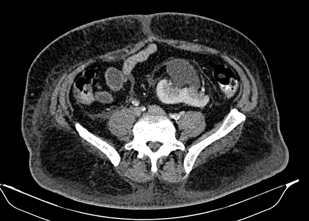
Left kidney is ectopic, lying in the pelvis at the pre-sacral region. It has short left ureter. The left renal artery and vein are single, where the left renal artery is arising from the left common iliac artery and the left renal vein is draining into the medial wall of inferior vena cava.
A small ureteric calculus noted within the left mid ureter, measuring 0.3cm in diameter, resulting in proximal hydroureter and moderate degree of left hydronephrosis. Significant degree of left perinephric and periureteric fat stranding indicates on-going inflammatory and infective process.
Small renal cortical cyst at the left upper pole. No kidney seen at the left renal bed.
Normal right kidney in position and size.
Hypodense area at the inferior pole of spleen which can represent splenic infarct or collection.
Bilateral moderate pleural effusion. A small round hypodense lesion within the left ventricle which raises suspicion of intra-cardiac thrombus.
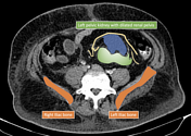
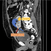
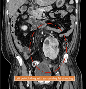
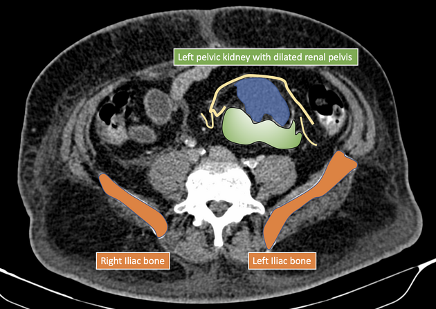
Annotated images showed the left ureteric calculus with obstructive uropathy as well as the left pelvic kidney.
Case Discussion
Pelvic kidneys are an uncommon variant and result from failed migration. The vascular supply of pelvic kidneys is highly variable. Ectopic kidney is prone for urinary tract infection, trauma and calculus disease compared to normally positioned kidney.


 Unable to process the form. Check for errors and try again.