
Presentation
Infant presenting with 24 hour history of urinary retention.
Patient Data
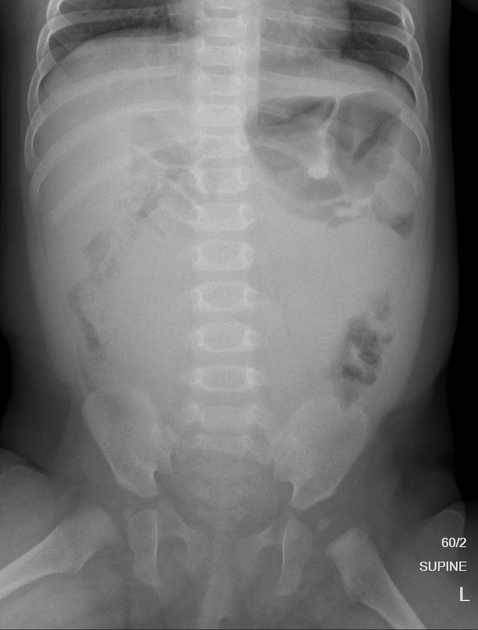
Supine abdominal radiograph demonstrates a marked paucity of gas centrally and in the pelvis with displacement of bowel loops laterally. Appearances are suspicious for a pelvic mass and/or distended urinary bladder.
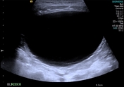
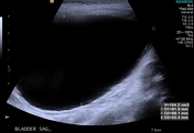
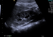
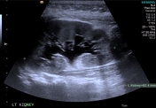
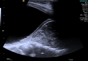
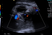
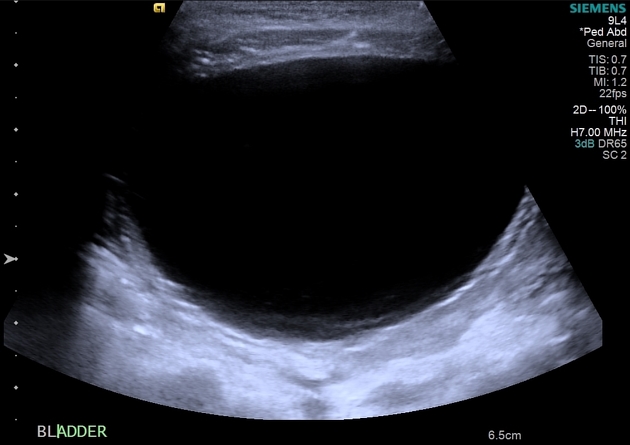
Abdominal ultrasound confirms a large volume urinary bladder without abnormal wall thickening and no intrinsic bladder mass. There is mild bilateral hydronephrosis, left greater than right. A soft tissue mass with internal vascularity is demonstrated posteroinferiorly in the pelvis, inferior to the bladder and anterior to the sacrum. The gynecological organs and rectum cannot be demonstrated.
Appearances are consistent with bladder outflow obstruction secondary to the pelvis mass.
An MRI was arranged for further assessment of the mass.
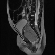
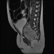
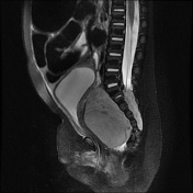
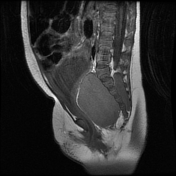
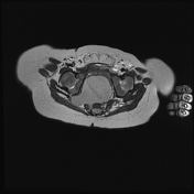

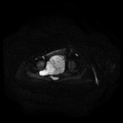
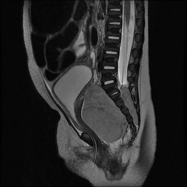
MRI pelvis confirms the presence of a well defined soft tissue mass (low T1 signal and iso-high intensity T2 signal) anterior to the sacrum with extension through the right sacral neural foramina into the spinal canal and extension laterally into the right greater sciatic notch. Despite encasing the sacrum, there is no intrinsic marrow signal change to suggest involvement. There is a clear fat plane between the mass and anteriorly sited uterus/vagina and urinary bladder. The mass demonstrates diffusion restriction. There was minimal heterogeneous enhancement following contrast administration.
Imaging of the remainder of the abdomen confirmed mild bilateral hydronephrosis. Normal appearances of the adrenal glands. No lymphadenopathy.
Conclusion:
Parasacral mass with intraspinal extension and invagination along neural pathways is consistent with a pelvis neuroblastoma.
There are no imaging features of sacrococcygeal teratoma.
Case Discussion
A presacral mass in an infant will more commonly represent a pelvic rhabdomyosarcoma or a sacrococcygeal teratoma than a neuroblastoma. However, the imaging characteristics in this case are typical for a neuroblastoma and the diagnosis was subsequently confirmed on open biopsy.


 Unable to process the form. Check for errors and try again.