
Download
Info

Normal reference images of the pelvis in a nine month old infant. Note that the image is normal but rotated, accounting for the asymmetry.
Download
Info
Normal reference images of the pelvis in an eleven month old infant.
Download
Info

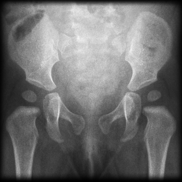
Normal reference images of the pelvis in an older child.
Case Discussion
The bony pelvis is formed by the sacrum and coccyx and a pair of hip bones ("ossa coxae"), which are part of the appendicular skeleton. Its primary function is the transmission of forces from the axial skeleton to the lower limbs as well as supporting the pelvic viscera.


 Unable to process the form. Check for errors and try again.