
Presentation
Incidental finding, asymptomatic.
Patient Data
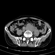

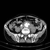
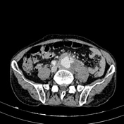
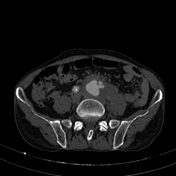
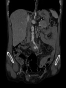
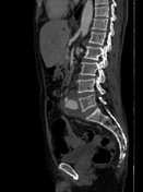
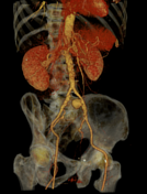

Calcification with atherosclerosis of the abdominal aorta and bilateral iliac arteries causes irregular arterial wall thickening, suggesting penetrating atherosclerotic ulcers (PAU). The left common iliac artery shows contrast extravasation forming a pseudoaneurysm measuring approximately 44 x 34 x 45 mm, with partial intraluminal thrombosis and mild surrounding fat infiltration.
A right inguinal hernia was also observed, containing a loop of small intestine, with no complications noted.
Surgical report:
During the operation, a pseudoaneurysm of the left common iliac artery, approximately 8 cm in diameter, was observed with a thin anterior wall. Upon incision of the aneurysmal sac, a small amount of dark brown turbid fluid was found. The sac wall was filled with atherosclerotic material, friable tissue, and the aneurysmal sac had ruptured into the retroperitoneum. Reconstruction of the abdominal aorta and bilateral common iliac arteries was performed using a Y-shaped 14-7 mm synthetic graft. The anastomoses were checked, ensuring patency and no bleeding was observed.
Case Discussion
The imaging and intraoperative findings are consistent with a penetrating atherosclerotic ulcer (PAU) of the common iliac artery complicated by pseudoaneurysm formation.
The patient underwent surgery to replace the infrarenal abdominal aorta and iliac arteries with a synthetic vascular graft. Postoperatively, the patient's condition stabilized, and they were discharged after a short recovery period.
PAU, along with aortic dissection and intramural hematoma, constitutes the spectrum of acute aortic syndrome.
Case co-author: Consultant specialist Tran Quyet Thang, Military Hospital 175, Vietnam.


 Unable to process the form. Check for errors and try again.