
Presentation
Two weeks history of right upper abdominal and epigastric pain.
Patient Data
Age: 60 years
Gender: Male
From the case:
Perforated duodenal ulcer with abscess formation

Download
Info
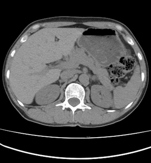
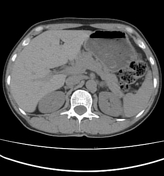
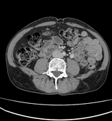
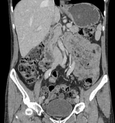
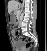
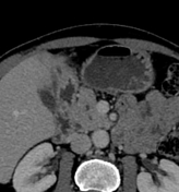
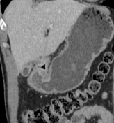
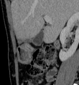
There is a small fluid collection within the segment IV of the liver with mild peripheral enhancement in continuity with a fistulous tract extending from the wall of the first part of the duodenum (well-visualized on zoomed images).
Case Discussion
CT findings are most consistent with a perforated duodenal ulcer with a small abscess formation within segment IV of the liver which was confirmed at surgery.


 Unable to process the form. Check for errors and try again.