
Presentation
Non specific pelvic pain.
Patient Data
Age: 75 years
Gender: Female
From the case:
Perineal hernia - posterior


Show annotations
Download
Info
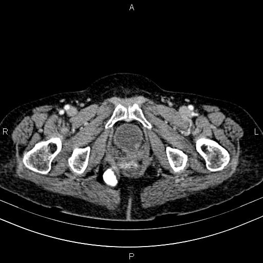
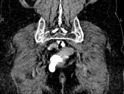
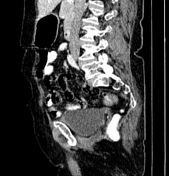
There is herniation of the rectum through the levator ani muscle into the right ischioanal fossa, without signs of bowel wall ischemia. The surrounding fat is intact.
Case Discussion
This case demonstrates a perineal hernia, also known as levator or pudendal hernia, a rare pelvic hernias, occurring through a defect in the pelvic floor musculature. Anterior hernias demonstrate herniation of the bowel through the urogenital diaphragm. Posterior perineal hernias are seen on CT as protrusion of loops of the sigmoid colon or rectum into the ischioanal fossa, as in this case.


 Unable to process the form. Check for errors and try again.