
Presentation
Craggy subcutaneous mass in right lateral canthal region. Normal eye movements and painless, but increasing eyelid/cheek swelling. Biopsy performed and MRI arranged.
Patient Data
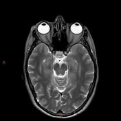

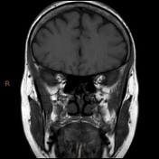
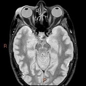
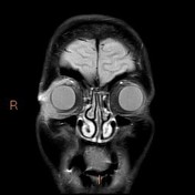
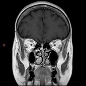
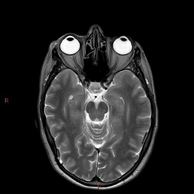
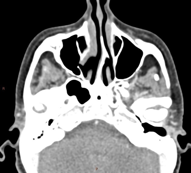
Ill-defined thickening and enhancement of pre and post-septal soft tissues in the right upper outer quadrant. Extraconal extension involving pre-aponeurotic fat and expected position of lacrimal gland (note absent lacrimal tissue on the left). Thin central fluid component anterior to lateral rectus insertion.
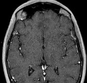
No globe, extraocular muscle, optic nerve or orbital apex involvement. No bone involvement. Normal intracranial appearances.
No periorbital mass three years earlier.
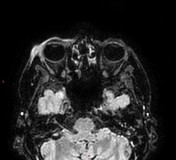
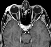
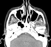
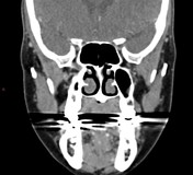
Note made of absent lacrimal glands bilaterally, as well as atrophic changes of parotid and submandibular glands.
Although imaging can be complicated by post-biopsy appearances, the differential here was most concerning for lymphoma - especially given the features of salivary gland atrophy (and a revealed history of known Sjogren syndrome - associated MALToma).
Inflammatory disorders including IgG4 disease and atypical infections also considered.
Subsequently biopsy results demonstrated non-caseating granulomatous appearance. No features of malignancy. Mild elevation of serum IgG, and normal serum ACE.
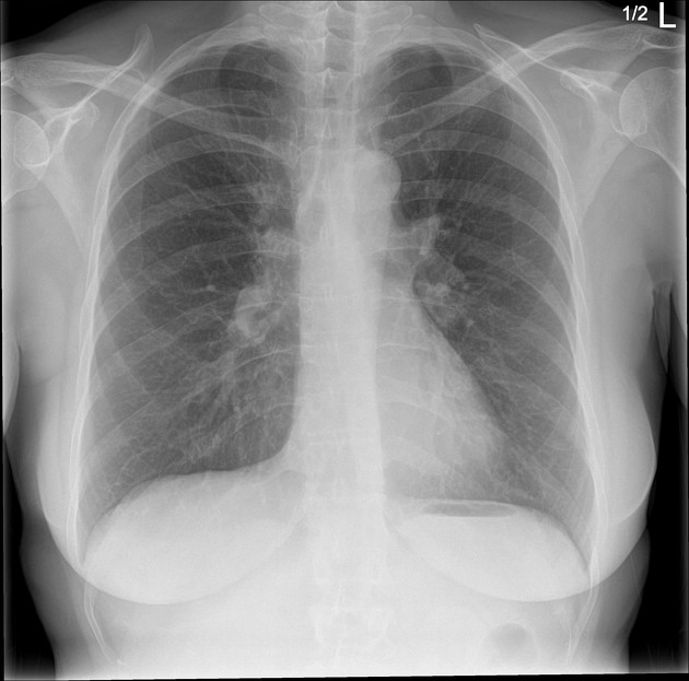
Bilateral hilar lymphadenopathy (compared with a historic CXR).
No pulmonary masses or pleural abnormality.
Case Discussion
An interesting case with multiple possible differentials but multisystem imaging features, pathological findings and good clinical response to steroids make the diagnosis of sarcoidosis the most likely etiology.
The additional findings associated with Sjogren syndrome, so far, appear to be coincidental.


 Unable to process the form. Check for errors and try again.