
Presentation
Pain for about three months without trauma.
Patient Data

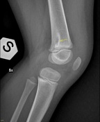
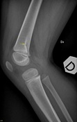
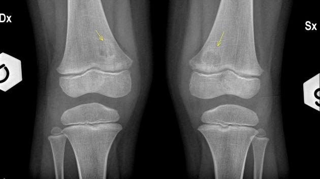
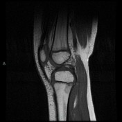
Knee frontal radiograph shows bilaterally a well-defined, eccentric lucency of the distal medial femoral metaphysis. Lateral radiograph demonstrates a focal cortical irregularity (yellow arrow) in the posterior aspect of the distal femoral metaphysis with adjacent periostitis.
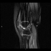

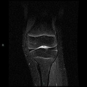
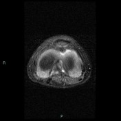

Axial MRI shows posteromedial cortical irregularity with associated periosteal reaction. Sagittal MRI localizes the defect to the femoral origin of the gastrocnemius muscle (medial head).
Case Discussion
Distal femoral metaphyseal irregularity (cortical desmoid) is an irregular cortical margin with an associated lucency found in the posteromedial aspect of the distal femoral metaphysis. It is thought to be an avulsion off the medial supracondylar ridge of the distal femur. These lesions may or may not be associated with pain. They are primarily found in young boys. The lesions are often bilateral.
It is a tumor like lesion developed by repeated micro-trauma of the origin of distal fibers of adductor magnus and aponeurotic origin of the medial head of gastrocnemius.


 Unable to process the form. Check for errors and try again.