Presentation
Diffuse abdominal pain and distension.
Patient Data
![](https://prod-images-static.radiopaedia.org/images/60569958/793ebaadf07fbc162f543783e407402728049bc74e6f600d6902feb778231601_thumb.jpeg)
![This study is a stack](/packs/stack-YQKLCKBI.gif)
![](https://prod-images-static.radiopaedia.org/images/60570275/b3c2b31d18824ccf4427f8bdf311b0d7219f2cfdc069f09f51ca7c53d0e34663_thumb.jpeg)
![This study is a stack](/packs/stack-YQKLCKBI.gif)
![](https://prod-images-static.radiopaedia.org/images/60570431/3b438801dd142a4e5dadc189d2d86c3d3e80293a1b40e0f3b71a22dafe3afeeb_thumb.jpeg)
![This study is a stack](/packs/stack-YQKLCKBI.gif)
![](https://prod-images-static.radiopaedia.org/images/60570492/5f3591ffbb0860fd090fc03c79a05ca7cd2344816dd2949a20a3b6d8e6582c09_thumb.jpeg)
![This study is a stack](/packs/stack-YQKLCKBI.gif)
![](https://prod-images-static.radiopaedia.org/images/60570533/78d389bd57b15615be6f9c15f8e23737a70c8d2f68e9fe5a534cc848e1dd7b3d_thumb.jpeg)
![This study is a stack](/packs/stack-YQKLCKBI.gif)
![](https://prod-images-static.radiopaedia.org/images/60570562/9596b996f4e6baa2e7f77a8d7859ee61c1efa9e1e2e3dda43d8fdf260496d9c7_thumb.jpeg)
![This study is a stack](/packs/stack-YQKLCKBI.gif)
![](https://prod-images-static.radiopaedia.org/images/60569958/793ebaadf07fbc162f543783e407402728049bc74e6f600d6902feb778231601_big_gallery.jpeg)
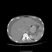
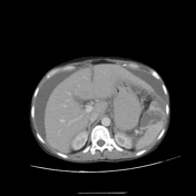
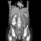
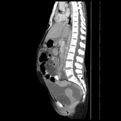
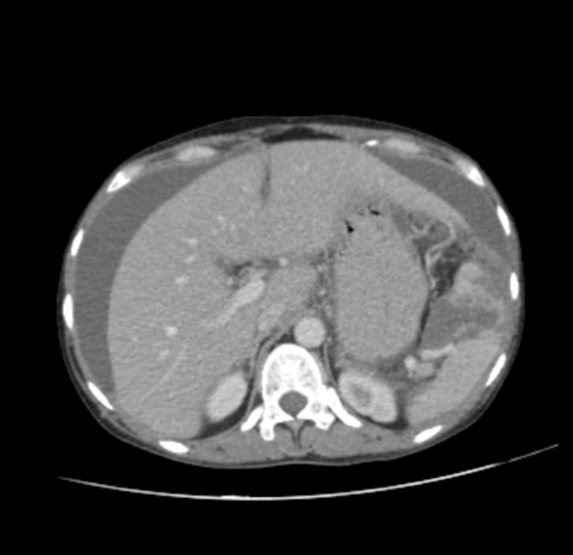
Moderate abdominal and pelvic ascites, associated with diffuse smooth peritoneal thickening and nodular omental thickening.
Multiple enlarged mesenteric lymph nodes showing hypodense cystic center.
At the splenic hilum, there are hypodense confluent lesions (possibly amalgamated nodes) infiltrating the splenic parenchyma.
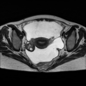
Both ovaries appear bulky yet with no masses.
Lower chest cuts showed bilateral pleural effusions and multiple enlarged necrotic mediastinal lymph nodes.
Radiological findings (mainly splenic involvement) were suggestive of abdominal TB.
![](https://prod-images-static.radiopaedia.org/images/60571155/02ca9c877ffda072a85e73329455983423b1f4dad68030179b44966bea62f167_thumb.jpeg)
![This study is a stack](/packs/stack-YQKLCKBI.gif)
![](https://prod-images-static.radiopaedia.org/images/60571201/adf1d3e4334fdd37ca4c75c729c04ba4dcc35d24c076fbf3fae7d898d418f020_thumb.jpeg)
![This study is a stack](/packs/stack-YQKLCKBI.gif)
![](https://prod-images-static.radiopaedia.org/images/60571155/02ca9c877ffda072a85e73329455983423b1f4dad68030179b44966bea62f167_big_gallery.jpeg)
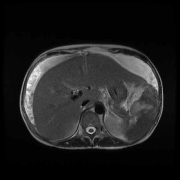
MRI confirmed the peritoneal and omental thickening, enlarged necrotic mesenteric and splenic hilar nodes with splenic involvement.
Right and left ovaries were of normal size and morphology showing prominent follicles, yet with no masses.
Case Discussion
Radiological findings include necrotic mediastinal and abdominal nodes, splenic involvement, and omental thickening as well as an absence of ovarian neoplasms on MRI. All those findings were suggestive of abdominal tuberculosis rather than peritoneal carcinomatosis. The diagnosis was confirmed by an omental biopsy and positive TB GeneXpert test. No malignancy.