
Presentation
Developmental delay with history of prematurity.
Patient Data
Age: 14 months
Gender: Female
From the case:
Periventricular leucomalacia (PVL) - end-stage


Download
Info
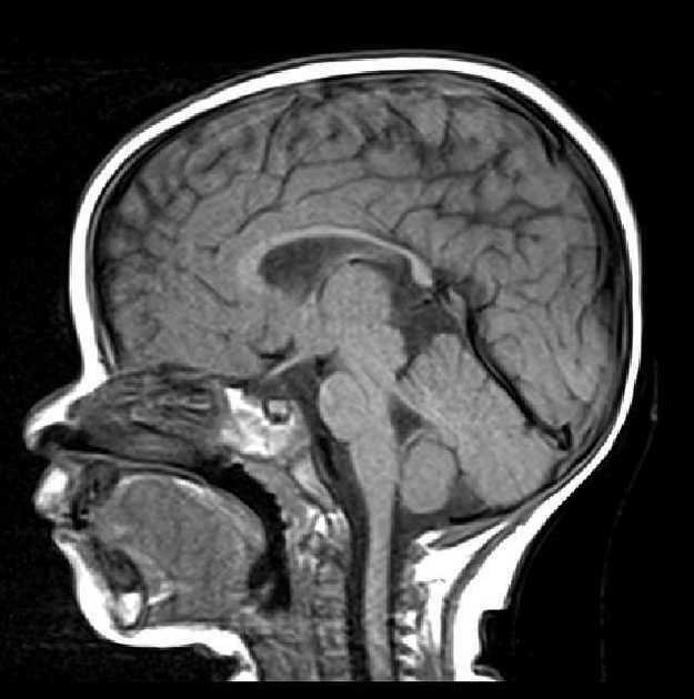
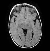
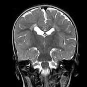
MRI sequences demonstrate:
- dilatation with irregular margins of the bodies and trigones of the lateral ventricles,
- loss of periventricular white matter with increased FLAIR and T2 signal containing small cystic formations
- thinning of the corpus callosum mainly of its corporeosplenial region
Case Discussion
MRI features of end-stage periventricular leucomalacia.


 Unable to process the form. Check for errors and try again.