
Presentation
Premature baby boy, 26 weeks GA
Patient Data
Age: Neonate
Gender: Male


Download
Info
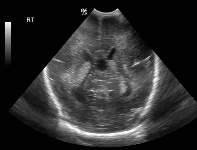
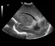
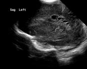
On 3 days old, there is bilateral periventricular echogenic area related to body of lateral ventricles, more prominent on left side.
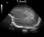
On 14 days old, partial cystic changes are seen within the periventricular lesions.
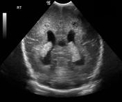
On 8 weeks old, obvious cystic changes are seen, larger on left side.
Features of periventricular leukomalacia (PVL).

Download
Info

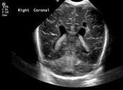
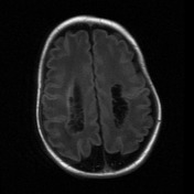
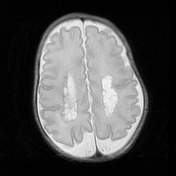
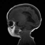
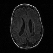
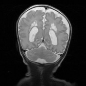
Bilateral cystic changes involving periventricular white matter along body of both lateral ventricles. Features of periventricular leukomalacia (PVL).
Case Discussion
Ultrasound and MRI studies show the progression of periventricular encephalomalacia.


 Unable to process the form. Check for errors and try again.