
Presentation
Follow up examination. The patient is asymptomatic at the time of the CT scan.
Patient Data
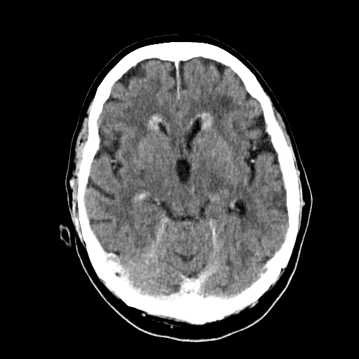
There is irregular soft tissue thickening along the ependymal surface of lateral venricles, specifically around the frontal and occipital horns.
The lateral ventricles show slight enlargement compared to the previous scan (not shown).
There is no evidence of a parenchymal mass, nor is there any abnormal enhancement of the pachymeninges or leptomeninges.
A brain MRI is needed for further investigation.

The patient was unfortunately lost to follow-up, and the MRI was performed three months after the initial CT scan.
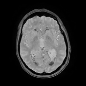
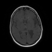
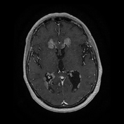
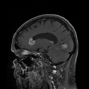
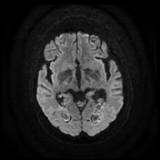
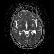
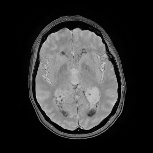
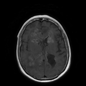
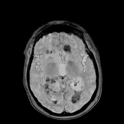
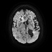
There is irregular and nodular soft tissue thickening along the ependymal surface of the lateral ventricles, which has progressed compared to the earlier CT scan. There are hyperintense areas on T1-weighted images, hypointense on gradient echo imaging with a heterogeneous appearance on diffusion weighted imaging (DWI), and some regions appearing hyperintense. After contrast injection (axial and sagittal), there is moderate and relatively homogeneous enhancement, along with some linear structures indicating intralesional small vessels.
Additionally, linear leptomeningeal hypointensities are observed on gradient echo images, particularly in the sylvian fissures, brainstem and cerebellum, consistent with superficial siderosis.
The lateral ventricles (including the temporal horns) and third ventricle appear more dilated compared to the previous CT scan.
The combination of hyperintensity on T1-weighted imaging (T1-WI) and hypointensity on T2*-weighted imaging (T2*-WI) suggests the presence of melanin. The patient had previously been treated for melanoma in the leg, which had been in complete remission for one year. Unfortunately, the disease has recurred, but it is now localized only in the brain, specifically along the ependymal surface of the lateral ventricles.

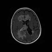
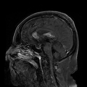
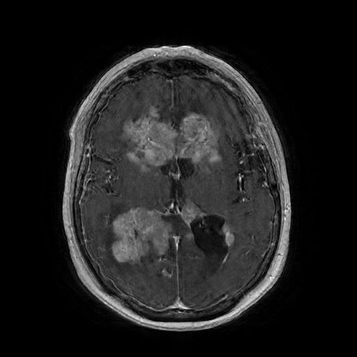
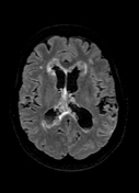
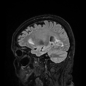
Unfortunately, the patient did not respond to treatment. The brain MRI shows progression of nodular ependymal soft tissue thickening, characterized by diffuse hyperintensity on T1-weighted imaging and hypointensity on T2*-weighted images. The size of the ventricles has also increased.
Additionally, T2*-hypointense layering is observed in the occipital horns of the lateral ventricles and in the recess of the fourth ventricle, which is consistent with hemorrhage. Superficial siderosis appears to be widespread, affecting both infratentorial and supratentorial regions.
Furthermore, confluent white matter hyperintense lesions are noted on FLAIR sequences in the supratentorial region, indicating vasogenic edema around the lesions and some degree of transependymal resorption.
Finally, there is effacement of the convexity sulci due to increased intracranial pressure.
Case Discussion
The patient had a history of leg melanoma with metastasis to the tibia, diagnosed and treated two years prior. A whole-body CT scan revealed no additional abnormalities, and the patient did not have a history of other cancers.
This case highlights an unusual pattern of melanoma recurrence in the brain along the ventricles with subependymal spread. Although uncommon, subependymal metastasis has been reported in lung cancer and very rarely in melanoma. The pathophysiology of this type of spread is not well understood.
Unfortunately for the patient, treatment was delayed by three months, and she did not respond well to systemic therapy. As a result, her condition worsened, and follow-up MRI scans indicated progressive disease.
The primary focus was identifying intralesional hyperintensities on T1-weighted imaging, hypointensities on gradient echo imaging, and superficial siderosis, which can be attributed to melanin content and related blood products.


 Unable to process the form. Check for errors and try again.