
Presentation
Right breast lump of 1 year duration.
Patient Data


Heterogeneously dense breast parenchyma (ACR c). A large encapsulated circumscribed mixed mainly isodense lesion is seen at the right lower central region.
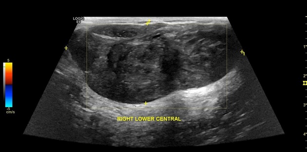
A large well-defined oval shaped hypoechoic lesion is seen extending from the right 5 -8 o'clock measuring about 6 x 3 cm along its maximum dimensions showing mild cystic changes.
Case Discussion
Well circumscribed large breast mass with the differential diagnosis including giant fibroadenoma and a phyllodes tumor.
Patient`s older age with rapid progressive clinical course together with mammographic findings of well-circumscribed high-density mass and sonographic findings of heterogeneous internal echogenicity, the presence of cystic components and posterior enhancement favors the diagnosis of phyllodes tumor.
Right breast mass WLE histology
Cut sections showing whitish-pink material with a focal reddish area (1.5x1.5 cm) and a cyst 0.5x0.5 cm in diameter.
Microscopy shows cellular stroma with increased cellularity in some areas, entangling scattered mature ducts with pushing borders. No evidence of tumor necrosis, cytologic atypia, invasive margin, heterogenous elements. Focal fibro-adenomatous and fibrocystic changes are observed. Safety margin is free of tumor extension. Ki67 labeling index revealed positivity in about 10% of stromal cells, thus the case is consistent with Phyllodes tumor.
Final diagnosis phyllodes tumor.


 Unable to process the form. Check for errors and try again.