
Presentation
Headache, mild distal weakness and acute hyperreflexia.
Patient Data


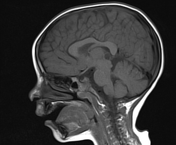
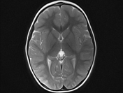
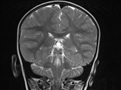

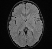
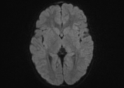
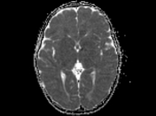
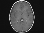
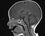
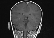
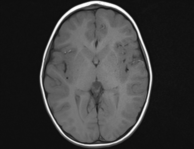
Pineal gland cyst is noted measuring 1.5 x 1 x 1.1 cm in dimensions. It is displaying CSF-like clear fluid intensities along all MRI sequences, no diffusion restriction and a thin rim of enhancement. It is indenting the upper part of the tectal plate of midbrain compressing the superior colliculi. It is slightly indenting the posteromedial aspect of both thalami. It is smoothly stretching the fine vascular structures of the pineal region.
Case Discussion
MRI study shows a well-defined pineal cyst, no other findings are noted that could be a possible cause for the presenting neurological manifestations. A pineal cyst is always asymptomatic and incidental finding, especially if less than 1 cm. if large or have atypical features, they cannot be distinguished from cystic tumors.


 Unable to process the form. Check for errors and try again.