
Presentation
Incidental finding on CT brain obtained for minor head injury. For further assessment.
Patient Data
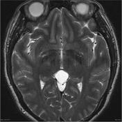


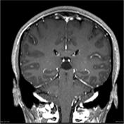
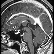
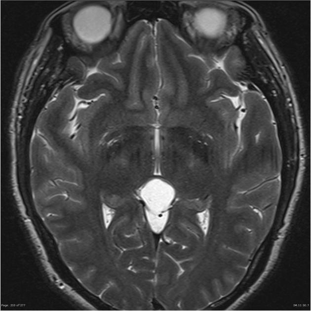
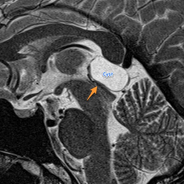
A well-circumscribed non-enhancing ovoid pineal mass measures 2.0 x 1.7 x 1.1 cm and distorts the upper tectum, mildly narrowing the upper aqueduct, with no associated signal abnormality or obstructive hydrocephalus.
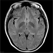
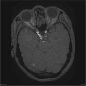
Note is made of a right trigeminal artery, which is moderately tortuous, but without overt sacular aneurysm. Corresponding hypoplasia of the basilar artery proximal to the trigeminal artery is also shown.

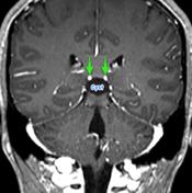
Pineal cyst compresses the superior colliculus (orange arrow) and splays and displaces the internal cerebral veins (green arrows) superiorly.
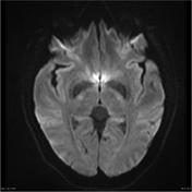
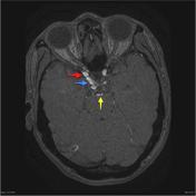
MRA demonstrates an aberrant trigeminal artery (blue arrow) arising from the large right internal carotid artery (red arrow) and passing posteriorly in an upwardly convex loop to the terminal basilar artery (yellow arrow).
Case Discussion
This case illustrates two incidental findings, a pineal cyst and a trigeminal artery. Pineal cysts are very common and are the cause of significant MRI followup examinations and stress for patients. Trigeminal arteries are rare, and the cause of stress for radiology residents / registrars, preparing to sit exams. Overall, the world would be a better place if neither entity existed.


 Unable to process the form. Check for errors and try again.