
Presentation
Increasing headaches.
Patient Data


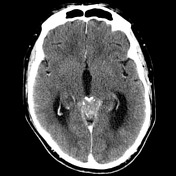
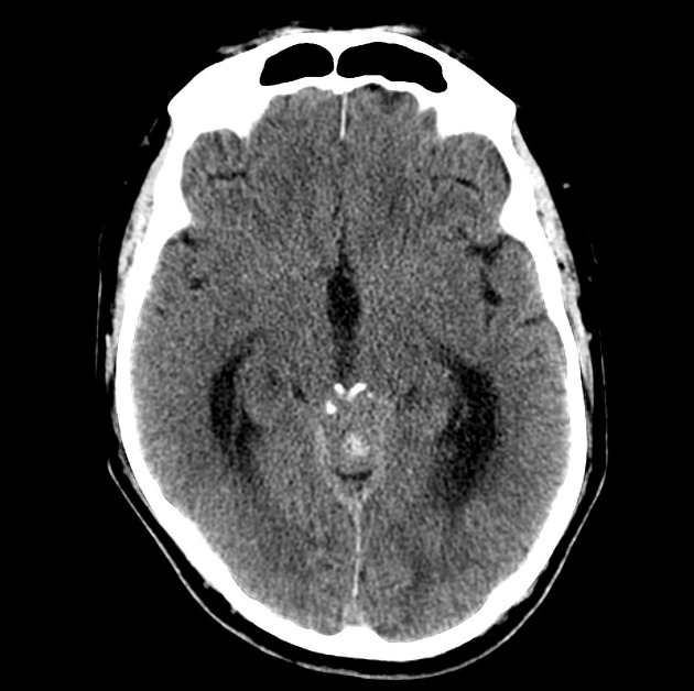
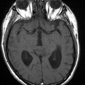
Pre-contrast scans showed moderate internal obstructive hydrocephalus, due to a large, partially calcified and dense mass, which appears to be centered upon the pineal gland. The quadrigeminal plate appears anteriorly displaced and the aqueduct obliterated. It does not appear to have an intimate relationship with the tentorium.
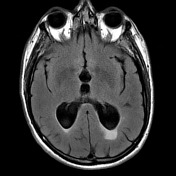
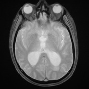
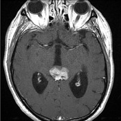
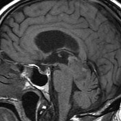

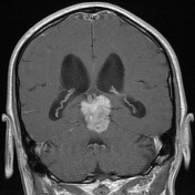
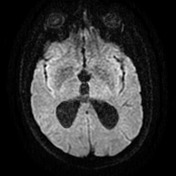

The irregular heterogenous enhancing pineal mass demonstrates several tiny cystic foci and eccentric coarse calcifications There is moderate mass effect on the adjacent tectum and vermis, with loss of definition and possible parenchymal invasion on the left.
There is associated aqueduct compression, with moderate hydrocephalus and hand and or signal abnormality.
Incidental note is made of minor paranasal sinus disease and polypoidal thickening at the posterior margin of the left inferior turbinate.

The patient went on to have a craniotomy and excision of the mass.
Histology
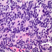
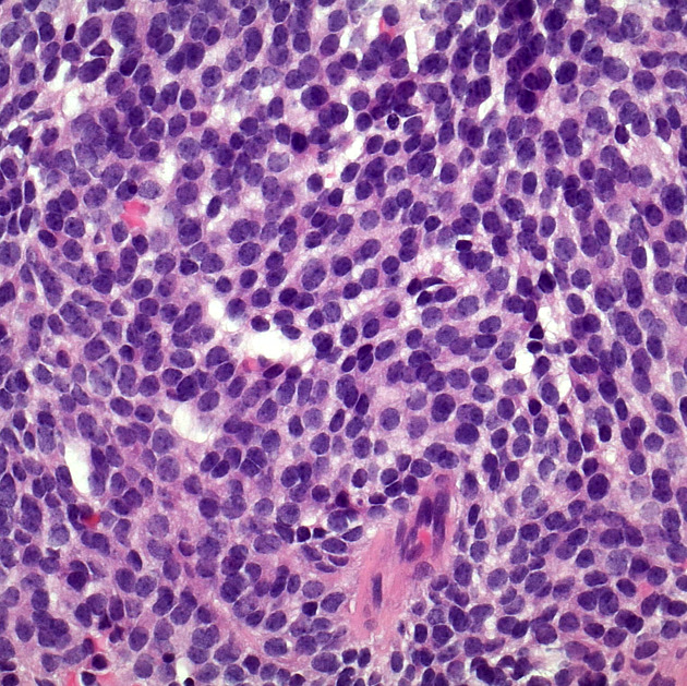
MICROSCOPIC DESCRIPTION: Paraffin sections show fragments of a densely hypercellular tumor. Tumor cells have small round, oval and angulated hyperchromatic nuclei and delicate processes which show strong immunostaining for neurofilament protein (NFP). Tumor cells are arranged in diffuse sheets. Prominent Homer-Wright rosettes are noted in several areas. Scattered mitotic figures are identified. No areas of necrosis are seen. Tumor cells show strong granular perinuclear immunostaining for synaptophysin. No staining for GFAP or NeuN is seen. The features are of pineoblastoma. The Ki-67/MIB-1 labeling index is approximately 6%.
FINAL DIAGNOSIS: Pineoblastoma (WHO Grade IV)
Case Discussion
Although the imaging appearances are fairly typical of a pineoblastoma the demographics of the patient are quite unusual; the vast majority of pineoblastomas are seen in children.


 Unable to process the form. Check for errors and try again.