
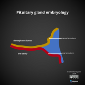


A simple diagram demonstrating the development of the pituitary gland from the oral ectoderm and neural ectoderm. Additional diagrams of Rathke cleft cyst and pharyngeal hypophysis.
Case Discussion
The pituitary gland is derived from two separate elements; the Rathke pouch of the oral ectoderm and the infundibular process (or infundibulum) of the neural ectoderm. These components later form the anterior and posterior lobes respectively.
The Rathke pouch appears at approximately 3-4 weeks first as an evagination of the oral ectoderm that later grows dorsally towards the infundibular process of the neural ectoderm. Expanding mesoderm subsequently constricts and pinches the neck of Rathke's pouch until it loses its connection with the oral ectoderm; remaining adjacent to the infundibular process.
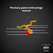
Cells of the anterior wall of Ranke's pouch later form the anterior lobe of the pituitary gland with a small extension surrounding the infundibular stem forming the pars tuberalis. The posterior wall of the Ranthke pouch forms the pars intermedia (of the anterior lobe).
The infundibular process forms the later infundibular stem (or pituitary stalk) and pars nervosa (posterior lobe of the pituitary gland).
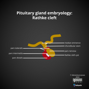
Should the Rathke cleft not completely regress and expand, the pouch remains as a remnant Rathke cleft cyst.
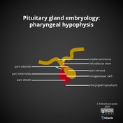
When the neck of the Rathke pouch is not completely obliterated, extrapituitary anterior pituitary tissue can be present in the roof of the nasopharynx. This is called a pharyngeal hypophysis.


 Unable to process the form. Check for errors and try again.