
Presentation
Patient had mild respiratory discomfort on climbing stairs.
Patient Data
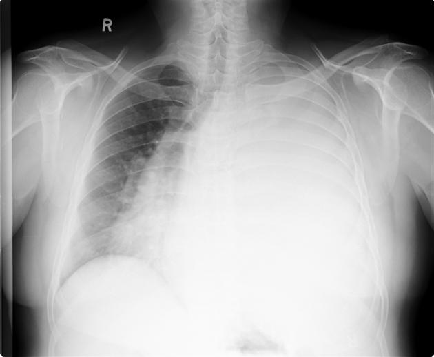
Opacification of the left hemithorax and right-to-left mediastinal shift with tracheal deviation.


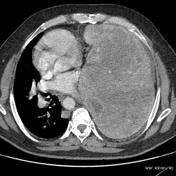

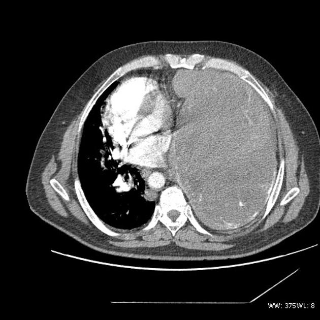
A very large well defined mass lesion of left pleura occupying the left hemithorax causing contralateral cardiomediastinal shift. The lesion appears hypovascular, with enhancing margins and internal lobulations, septations, and few calcific specks.
Minimal left pleural free fluid was also noted.
Dilated tortous engorged posterior paraaortic collateral vessels are seen.
Pulmonary angiographic phase showed no obvious vascularity from the pulmonary or bronchial arteries except for a solitary peripherally splayed prominent vessel at the basal region
Case Discussion
Findings are unchanged from CT that was performed six months prior.
CT biopsy showed a very acellular tumor, showing only collagen fibrils. A second biopsy was performed and revealed reticulin foci, with collagen and few spindle cells. Histopathology is consistent with pleural fibroma.


 Unable to process the form. Check for errors and try again.