
Presentation
Cough. Chest infection?
Patient Data
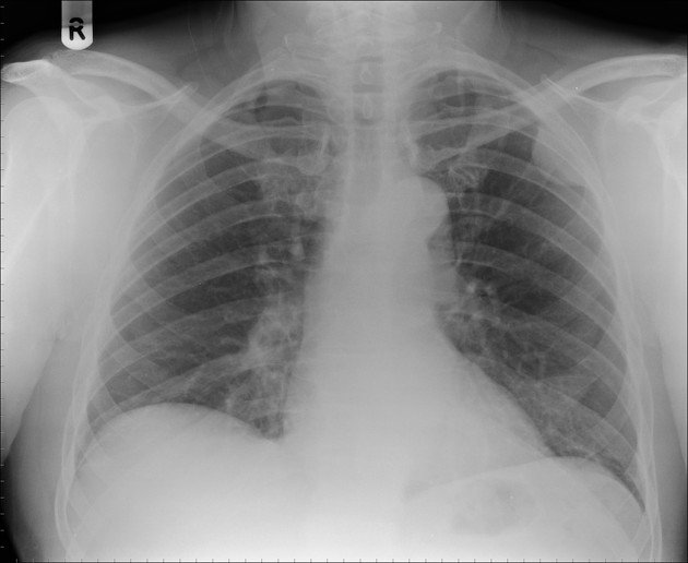
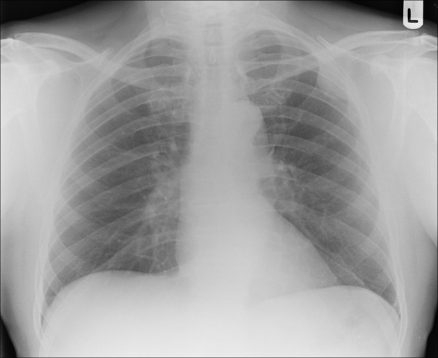
3cm well defined mass in the upper left hemithorax abutting the chest wall to which it forms an obtuse angle.
No bony destruction of the thoracic cage.
Heart size normal. Lungs clear.
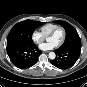

Well defined pleural based opacity at the left upper zone with a rather smooth surface, measuring 1.5 x 3.5 x 3.5 cm in size. The fat plane of the intercostal muscles is preserved, suggesting there is no evidence of invasion into the chest wall.
No hilar and no mediastinal mass lesion. The rest of the lungs are clear.
Fatty infiltration of the liver. No focal liver lesion. The spleen, adrenals, kidneys and pancreas are normal.
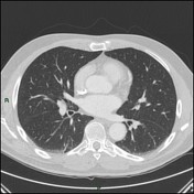
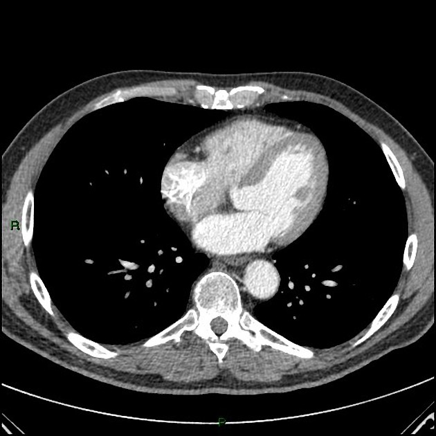
Unchanged left sided pleural based mass.
Heart size normal. Lungs clear.
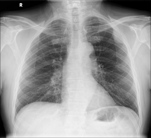
Unchanged left sided pleural based mass.
Heart size normal. Lungs clear.
Case Discussion
This is a chest wall-based mass, as evidenced by the obtuse angle it forms as opposed to an acute angle with the chest wall if it was pulmonary based. The CT confirmed this to be pleural based specifically.
Longitudinal studies over nearly a decade with no change in size or appearances indicates its benign nature and is almost certainly a pleural fibroma.


 Unable to process the form. Check for errors and try again.