
Presentation
Flu-like illness. Rash.
Patient Data
Age: 70 years
Gender: Male
From the case:
Pneumocystis jiroveci pneumonia

Download
Info
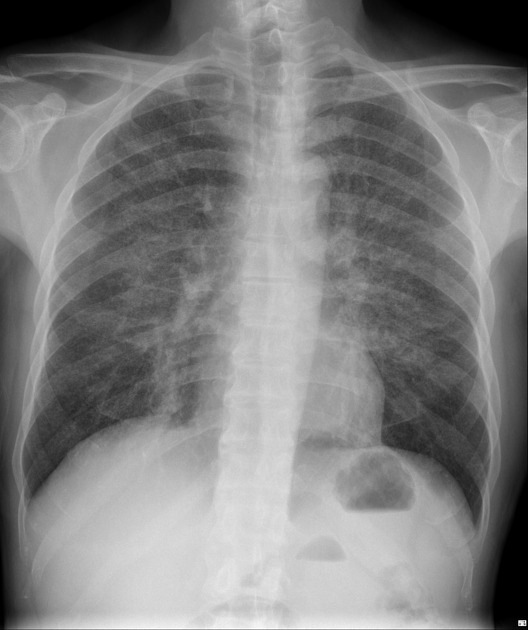
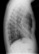
Diffuse interstitial opacities are seen throughout both lungs with a mid-to-upper zone predominance.
From the case:
Pneumocystis jiroveci pneumonia

Download
Info

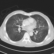
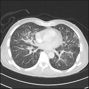
There are numerous small ill-defined lung nodules and groundglass opacities, particularly affecting the upper lobes. Atelectasis / fibrosis within the middle lobe medially.
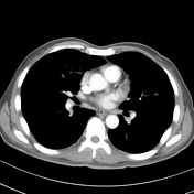
The mediastinum appears normal. No pericardial effusion.
The patient proceeded to bronchoscopy and bronchoalveolar lavage:
Pneumocystis jiroveci DNA by PCR: Detected
Case Discussion
Pneumocysitis pneumonia is an atypical infection which typically has perihilar ground glass opacity. The nodules in this case are somewhat atypical and a differential diagnosis of tuberculosis was given on imaging appearances.


 Unable to process the form. Check for errors and try again.