
Presentation
Cough and fever
Patient Data
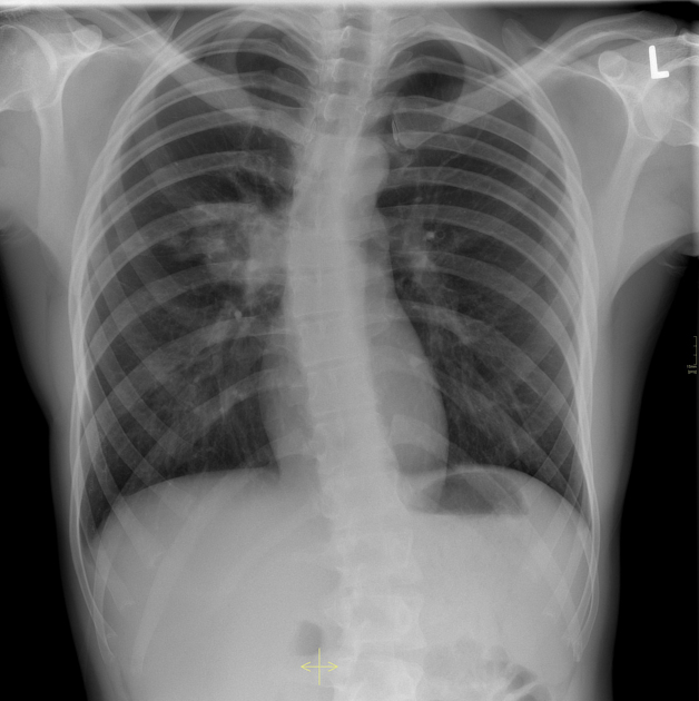
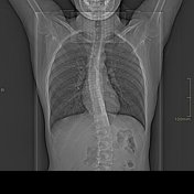
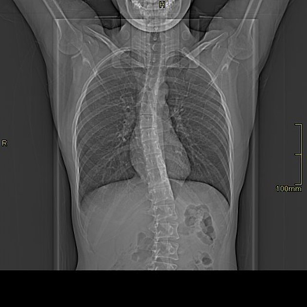
There is an opacity projecting in the right hilar/perihilar region with preserved hilar contour, indicating that the abnormality is not arising from the hilum. It can be located either anterior or posterior to the hilum.
Few nodular opacities are seen lateral to the forementioned opacity.

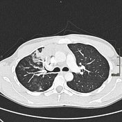


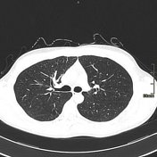
Contrast-enhanced CT scan of the chest performed on the same day shows consolidation in the anteromedial aspect of the right upper lobe with surrounding patchy air space disease, consistent with pneumonia.
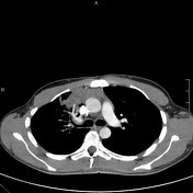
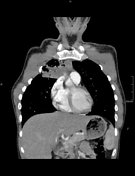
Follow up CT scan after treatment showed resolution of pneumonia.
Case Discussion
Hilum overlay sign is the ability to see the normal hilar outline through opacities projecting over the hilum, excluding pathologies being of hilar origin or involving the hilum.


 Unable to process the form. Check for errors and try again.