
Presentation
ICU. Intubated. Routine x-ray.
Patient Data
Age: 50 years
Gender: Male
From the case:
Pneumopericardium
Download
Info
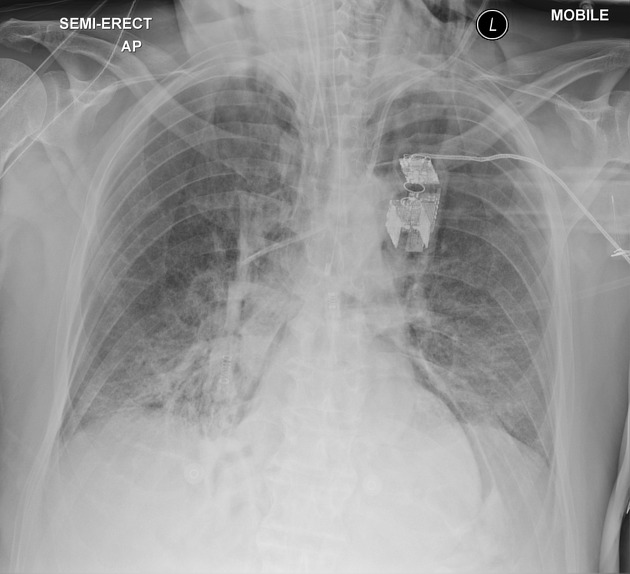
Patient is intubated with a left central venous catheter. NGT with tip in the mid-esophagus. Bibasal opacities. Lucency outlines the heart with a small volume of subcutaneous emphysema in the root of the neck.


Download
Info
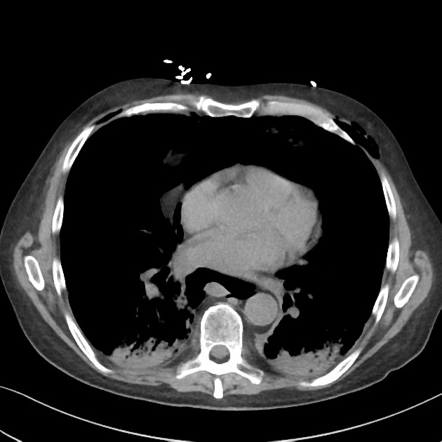
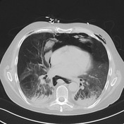
Extensive pneumomediastinum (including small volume pneumopericardium) and subcutaneous emphysema. No pneumothorax. Left psoas hematoma is partially imaged.
Case Discussion
The most likely cause is barotrauma from positive pressure ventilation. Marked worsening in the 8 hours between studies is also seen.


 Unable to process the form. Check for errors and try again.