
Presentation
Acutely unwell with respiratory compromise requiring intubation and ventilation.
Patient Data
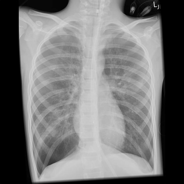
ET tube and NG tube in situ and in appropriate positions.
The lungs are hyperexpanded with flattening of the diaphragms. Appearances suggest background of asthma. At both apices, the lung edge can be seen with no lung markings peripherally. Features here are of small bilateral pneumothoraces.
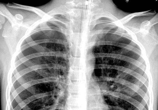
With a bit of windowing and zoom, both the apical pneumothoraces becomes much more obvious.
Case Discussion
The lungs are hyperexpanded and in a young patient, this should trigger thoughts of asthma. With asthmatic patients, look for a cause of their exacerbation (e.g. viral chest infection), but also look for secondary complications (e.g. pneumothorax, pneumomediastinum, surgical emphysema).
In the case of pneumothorax, use a good monitor, good viewing conditions and window to give yourself the best chance of finding it. Pneumothoraces are most likely to be apical in these patients, so give it a bit of zoom too.
Pneumothoraces are more common in asthmatics, but may also be the result of over-ventilation, either by hand-bagging or mechanical-ventilation.


 Unable to process the form. Check for errors and try again.