Presentation
Headache, lower limb weakness and visual disturbance
Patient Data
![](https://prod-images-static.radiopaedia.org/images/30843239/f6fb84d3f1da545e8ad37535b304dc_thumb.jpeg)
![This study is a stack](/packs/stack-YQKLCKBI.gif)
![](https://prod-images-static.radiopaedia.org/images/30842449/b126a74157c9daa21ceacf2923c898_thumb.jpeg)
![This study is a stack](/packs/stack-YQKLCKBI.gif)
![](https://prod-images-static.radiopaedia.org/images/30843047/45ab44f1f6b94cfdb08010f4a3d340_thumb.jpeg)
![This study is a stack](/packs/stack-YQKLCKBI.gif)
![](https://prod-images-static.radiopaedia.org/images/30843431/e1545a95efd39e50b5099d8d31d83c_thumb.jpeg)
![This study is a stack](/packs/stack-YQKLCKBI.gif)
![](https://prod-images-static.radiopaedia.org/images/30842247/c495e6c501b2fa066952f0c09e2b48_thumb.jpeg)
![This study is a stack](/packs/stack-YQKLCKBI.gif)
![](https://prod-images-static.radiopaedia.org/images/30842696/1d9d8d5a2b3fa0da8d7fe0556dfe64_thumb.jpeg)
![This study is a stack](/packs/stack-YQKLCKBI.gif)
![](https://prod-images-static.radiopaedia.org/images/30843861/de4f85dffdc8fba45664e3ec8af8b0_thumb.jpeg)
![This study is a stack](/packs/stack-YQKLCKBI.gif)
![](https://prod-images-static.radiopaedia.org/images/30844025/da88f97621b49826cde241d88dc81b_thumb.jpeg)
![This study is a stack](/packs/stack-YQKLCKBI.gif)
![](https://prod-images-static.radiopaedia.org/images/30843649/dbbc3b024c6c203d3f9323db14b621_thumb.jpeg)
![This study is a stack](/packs/stack-YQKLCKBI.gif)
![](https://prod-images-static.radiopaedia.org/images/30844249/8098aa8b78abd8181e1c9f690b8017_thumb.jpeg)
![This study is a stack](/packs/stack-YQKLCKBI.gif)
![](https://prod-images-static.radiopaedia.org/images/30843239/f6fb84d3f1da545e8ad37535b304dc_big_gallery.jpeg)
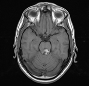
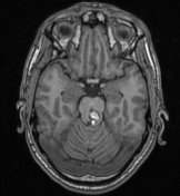
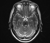
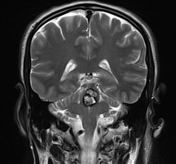
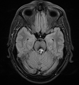
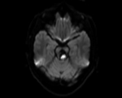
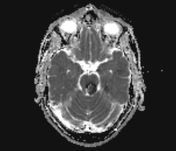
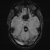
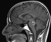
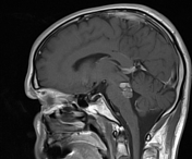
A rather defined (2.2 x 1.6 x 1.5 cm) "popcorn ball like" lesion is noted at posterior portion of the pons along the midline inclined to the left side and partially encroaching upon the inferior aspect of the midbrain. It displays T1 heterogeneous intensity with multiple foci of hyperintensity, T2 hyperintense signal, with hypointense hemosiderin ring and few fluid-fluid levels. Following intravenous contrast injection the lesion shows modest enhancement. On SWI the lesion shows marked blooming. No other similar lesions seen. Mild mass effect is exerted manifested as indentation of the anterior aspect of the fourth ventricle.
![](https://prod-images-static.radiopaedia.org/images/30842481/d4954a812cdea4463eec8486a842e8_thumb.jpeg)
![](https://prod-images-static.radiopaedia.org/images/30842950/96e5ed0d1b62150b62d6e83d577c18_thumb.jpeg)
![](https://prod-images-static.radiopaedia.org/images/30842958/0ce8d094997890d5c445520a6c5a4e_thumb.jpeg)
![](https://prod-images-static.radiopaedia.org/images/30842959/3213ec011eb74a514f8178b0fa78d7_thumb.jpeg)
![](https://prod-images-static.radiopaedia.org/images/30842976/2665a2dedec2de3b566a2a4a0ec573_thumb.jpeg)
![](https://prod-images-static.radiopaedia.org/images/30843014/a2561585001488d3a251246028bb7b_thumb.jpeg)
![](https://prod-images-static.radiopaedia.org/images/30843016/788cbda696f742b28f7559b79f70fd_thumb.jpeg)
![](https://prod-images-static.radiopaedia.org/images/30842978/ef535b85a840731747b1f34db4090e_thumb.jpeg)
![](https://prod-images-static.radiopaedia.org/images/30842977/3e2a634ee8bfdd3923a57cc0bee8b8_thumb.jpeg)
![](https://prod-images-static.radiopaedia.org/images/30842960/b37a52829400423a326de194b5b2d9_thumb.jpeg)
![](https://prod-images-static.radiopaedia.org/images/30842481/d4954a812cdea4463eec8486a842e8_big_gallery.jpeg)
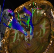
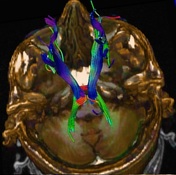
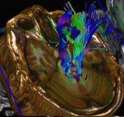
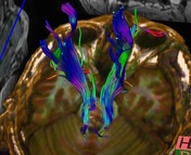
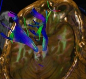
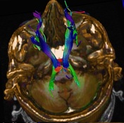
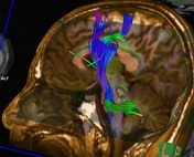
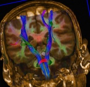
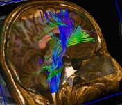
The corticospinal tracts are anterior to cavernoma with no displacement or fiber attenuation. The medial lemnisci on both sides are mildly displaced laterally with no fiber attenuation.
Case Discussion
MRI features are in favor of cavernous malformation "cavernoma" at the posterior aspect of the pons "Zabramski type 2". The corticospinal tracts are anterior to cavernoma with no displacement or attenuation of the fibers. The medial lemnisci on both sides are mildly displaced laterally with no attenuation of the fibers. MRI is the modality of choice, demonstrating a characteristic “popcorn” or "berry" appearance with a rim of signal loss due to hemosiderin.