
Post-intubation pneumomediastinum and pneumothorax - background COVID-19 pneumonia
Presentation
Increasing lethargy and flu-like symptoms. New oxygen requirement. Crackles on both bases
Patient Data
Day 1
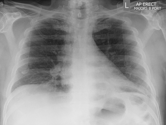
No collapse or consolidation. No pleural effusion.
Day 6
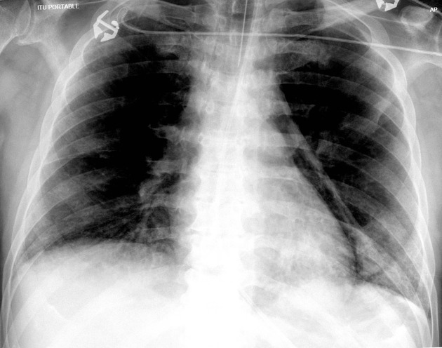
ETT and NG tube placed. New air-space shadowing with air bronchogram in left lower zone . New pneumomediastinum demonstrated.
Day 6
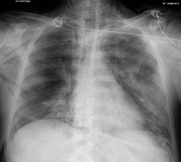
ETT and NG tube in place. Right-sided intercostal drain with bilateral small pneumothorax and pneumomediastinum with diffuse air space in both lungs typical for COVID.
Day 7
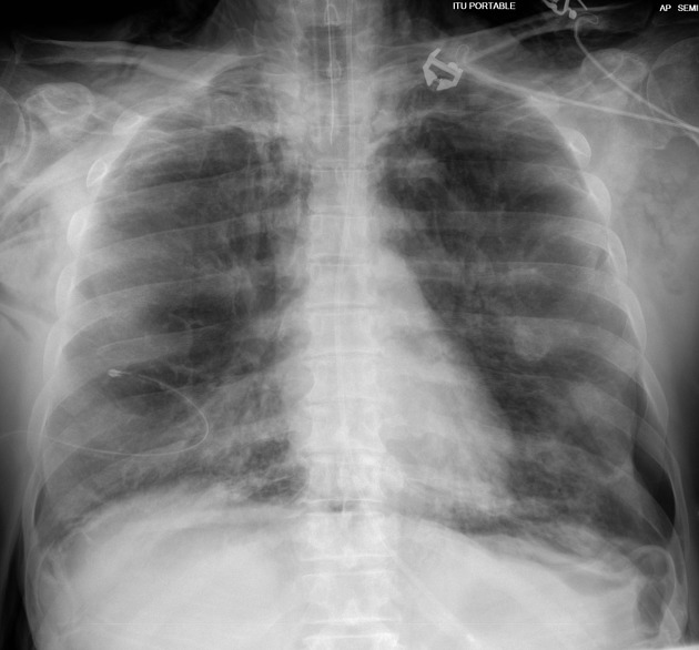
ETT, NG tube and right sided intercostal drain in place. Interval increase in size of left pneumothorax with persistent right pneumothorax and pneumomediastinum with diffuse air space in both lungs. No pleural effusion.
Case Discussion
This is a case of PCR proven SARS-CoV-2 with typical imaging findings of COVID-19 demonstrated on subsequent chest radiographs. Although acute respiratory distress syndrome (ARDS) does not affect lung compliance, patients can still present with pneumothorax as a complication of pneumomediastinum 1.
Thanks to Dr. Madava Djearaman (consultant radiologist), Dr. Shahid Hussain (consultant radiologist), Dr. Dilina Rajapakse and Dr. Haren Wijesinghe for their input.


 Unable to process the form. Check for errors and try again.