
Presentation
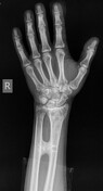
The patient presents to the emergency department with an index finger laceration. An x-ray series of the right hand was routinely taken to exclude the presence of any foreign objects. Upon inspection of the traumatic area, inability of the patient to perform pronosupination movement is observed.
Patient Data
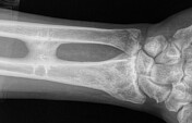
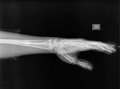

Incidentally, synostosis of the distal radius and ulna is noticed. History of right forearm trauma was reported.
The frontal x-ray of the right hand shows an osseous bridge connecting the distal third of the radius and ulnar diaphysis. The lucent lines indicate previous internal fixation of the transverse fractures of both the radius and ulna by using metal plates and screws.
Case Discussion
The incidence of post-traumatic radioulnar synostosis ranges from 0% to 9.4% among all forearm fractures, depending on factors such as the severity of the injury, the type of surgical intervention, and individual patient risk factors.
While the proximal region of the forearm is the most common site for synostosis to develop, it can also form in the middle and distal thirds of the forearm in rare cases, as seen in this patient.


 Unable to process the form. Check for errors and try again.