
Presentation
Recent onset of seizure. Previous medical history was unremarkable
Patient Data
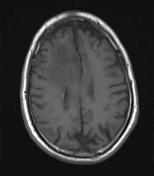

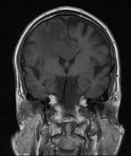
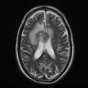
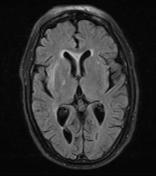
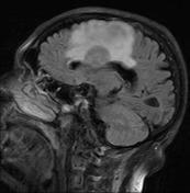
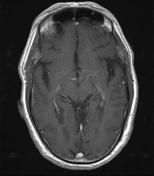
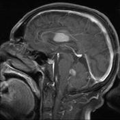
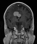
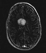
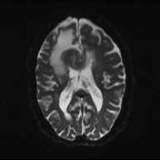
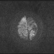
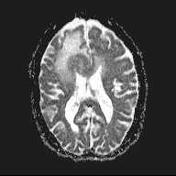
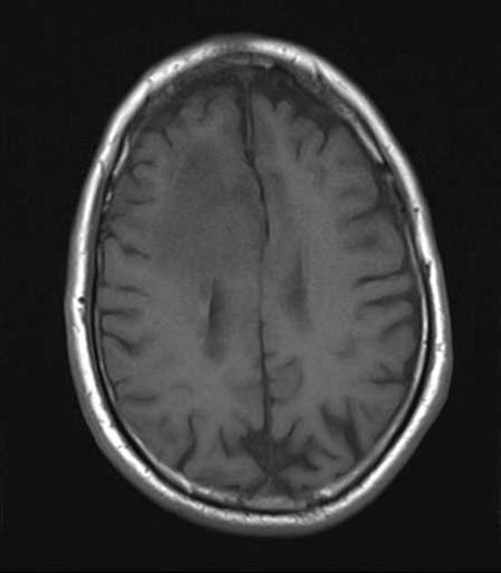
MRI shows a well defined intra axial space occupying lesion in the periventricular deep white matter of the right parietal lobe associated with extensive peripheral edema and mild midline shift.There is also another lesion in the peripheral aspects of the fourth ventricle.Following intravenous administration of gadolinium diffuse enhancement of the masses are noted.Note that the superior lesion also extends into the corpus callosum (best depicted in the post contrast coronal images) ,crossing the midline into the contralateral hemisphere and creating a butterfly pattern.In addition there is subependymal enhancemet along the lateral margin of the frontal horn of the right lateral ventricle in favor of subependymal tumor extension.
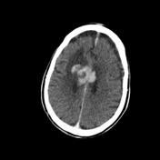
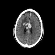

There is a hyperdense mass of the deep white matter of the parietal lobe. Postcontrast CT scan reveals intense enhancement.There is also evidence of craniotomy.
Case Discussion
The patient underwent craniotomy and biopsy was performed.
Histopathology revealed Malignant lymphoma (diffuse large cell type,high grade). Tumoral cells were positive for CD45,CD20 and negative for GFAP and EMA.


 Unable to process the form. Check for errors and try again.