
Presentation
Previous history of breast and mediastinal lymphoma. In regression after 6 cycles of chemotherapy.
Patient Data
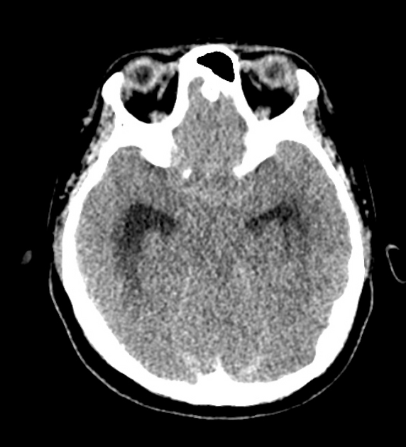
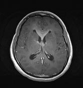
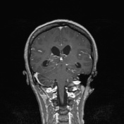
The temporal horns are disproportionate to the size of the rest of the ventricular system.
Brain parenchyma normal.
In the following 10 days the patients status changed from merely being ''out of sorts'' to confusion.


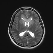

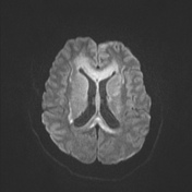
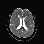
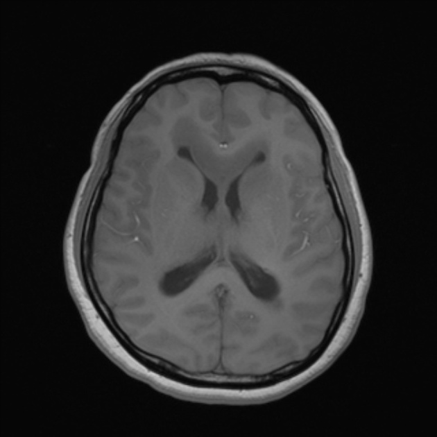
Enhancing infiltrative abnormality in the deep periventricular white matter along with the genu and body of the corpus callosum. The tissue also exhibits strong diffusion restriction.
Dilated temporal horns. The remainder of the ventricular system is normal.
Enhancing infiltrative mass involving the pituitary stalk.
Case Discussion
The appearances of those of primary CNS lymphoma.
Although a previous history of lymphoma is indicated no intercurrent disease was present ( in remission ) at the time of this study.
The pattern of disease involving the deep white matter and corpus callosum is more typical than the leptomeningeal disease typically identified in secondary CNS lymphoma.
The case also illustrates the striking superiority of MRI in imaging the brain - the extensive deep white matter disease is essentially unidentifiable on the CT brain.


 Unable to process the form. Check for errors and try again.