
Presentation
Headache with history of renal transplant
Patient Data

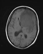
Hyperdense CT mass in a deep supratentorial location. There is extensive adjacent edema. On these images it is difficult to definitely distinguish an extra-axial lesion arising from the sphenoid wing, from an intraparenchymal lesions (the latter is favored).


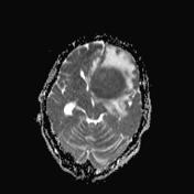
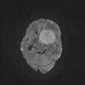
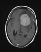
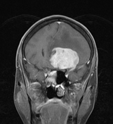
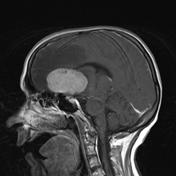

Solitary lesion is isointense on FLAIR and slightly hypointense on T1 images. Prominent restricted diffusion. Avid homogeneous enhancement is noted in post contrast images.
Case Discussion
The biopsy result was non Hodgkin's lymphoma.
The incidence of CNS lymphoma has increased in recent years due to the increased number of immunocompromised patients (organ transplant, HIV infection, etc.). In this clinical context, it represents primary CNS post-transplant lymphoproliferative disorder. In this case, the appearances are those seen in sporadic primary CNS lymphoma, without the features often present in immunocompromised patients (e.g. multiple lesions, central non-enhancement).


 Unable to process the form. Check for errors and try again.