
Presentation
Subacute history of weakness and altered sensorium
Patient Data
Note: This case has been tagged as "legacy" as it no longer meets image preparation and/or other case publication guidelines.

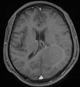
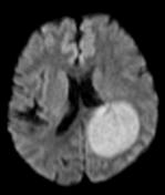

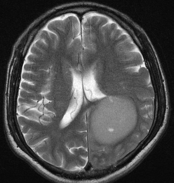
Left periventricular mass demonstrating a homogeneous, T2 iso- to hyperintense mass with patchy enhancement post gadolinium and prominent restricted diffusion.
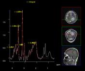
MR Spectroscopy shows a large Choline peak, reversed Cho/Cr and Lactate peaks. Prominent inositol peak is also seen.
Case Discussion
Left periventricular mass demonstrating a homogeneous, T2 iso- to hyperintense mass with patchy enhancement post gadolinium and prominent restricted diffusion.
An initial CT (not shown) demonstrated a homogeneously hyperdense mass.
The lack of prominent enhancement is atypical.
MR spectroscopy revealed: Large Choline peak with reversed choline:creatinine ratio. Prominent lactate peak is also seen. Prominent inositol peak is also seen.
Findings are nonetheless consistent with primary CNS lymphoma.


 Unable to process the form. Check for errors and try again.