
Presentation
Headache and increasing confusion. No sided weakness.
Patient Data


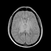
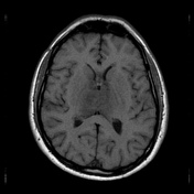
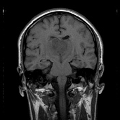
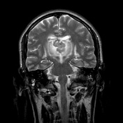
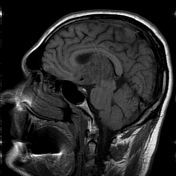

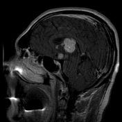
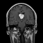
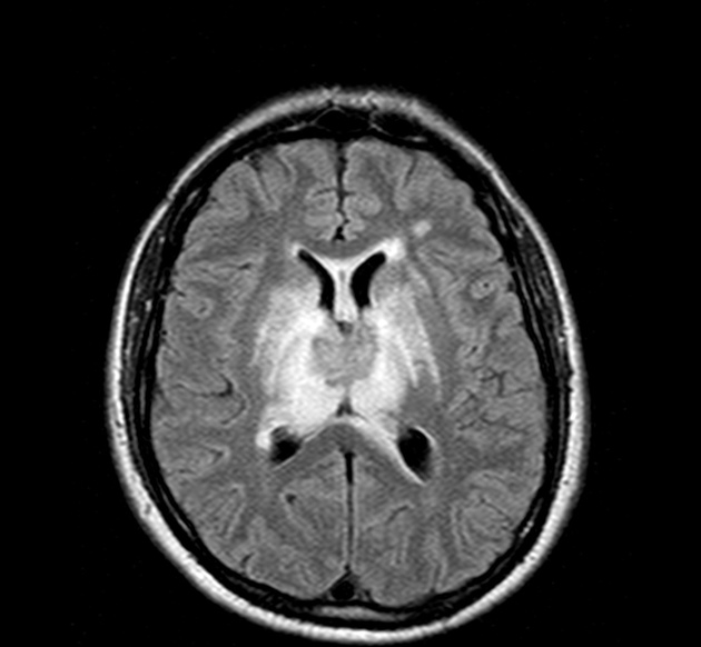
A large, round-shape, mass involving the territory from hypophysis to ventricles. After contrast a homogeneous impregnation is evident. Edema is present surrounding the lesion.
Case Discussion
Lymphoma of the CNS consists of 2 major subtypes: secondary CNS involvement by systemic lymphoma (the most common) and PCNSL, in which the lymphoma is restricted to the brain, leptomeninges, spinal cord, or eyes, without evidence of it outside the CNS at primary diagnosis.
Primary central nervous system lymphoma (PCNSL) is a rare disease, accounting for 6% of all intracranial malignant tumors and 1-2% of all lymphomas.
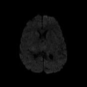
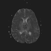
Single or multiple periventricular and/or superficial contrast-enhancing lesions are characteristic of parenchymal CNS lymphoma.
CT and MR imaging techniques and, recently, metabolic imaging have demonstrated characteristic findings in CNS lymphoma, aiding in its differentiation from other CNS lesions.
A biopsy diagnosed PCNSL; the patient is dead two years after diagnosis, despite chemotherapy and radiotherapy treatment.


 Unable to process the form. Check for errors and try again.