
Note: This case has been tagged as "legacy" as it no longer meets image preparation and/or other case publication guidelines.
From the case:
Primary CNS lymphoma


Download
Info
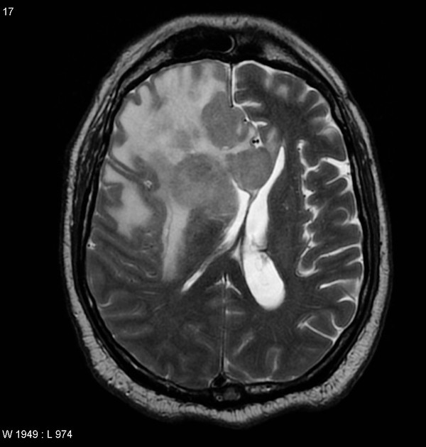
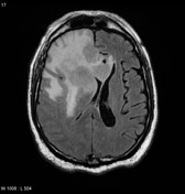
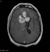
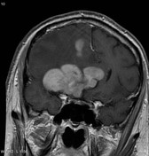
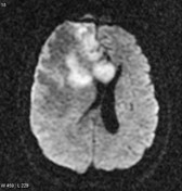
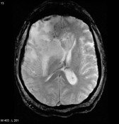
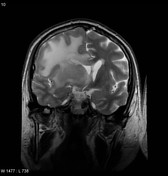
Selected images demonstrating a large multilobulated mass centered in the right frontal lobe. It homogeneously enhances and has somewhat lower T2 signal and prominent restricted diffusion. It extends to involve the caudate than thus the periventricular area. There is significant mass effect.
Case Discussion
This patient went on to have a biopsy that confirmed the diagnosis of primary CNS lymphoma.


 Unable to process the form. Check for errors and try again.