
Presentation
Skull radiograph for shunt valve settings. Ventriculoperitoneal shunt inserted for normal pressure hydrocephalus.
Patient Data
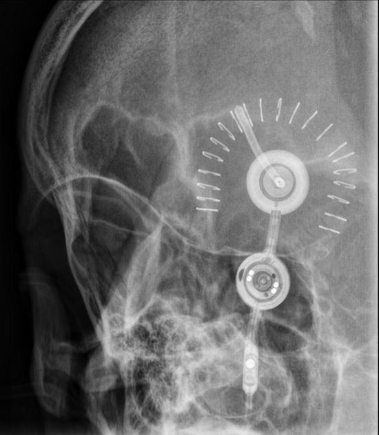
Ventriculoperitoneal shunt valve in place.
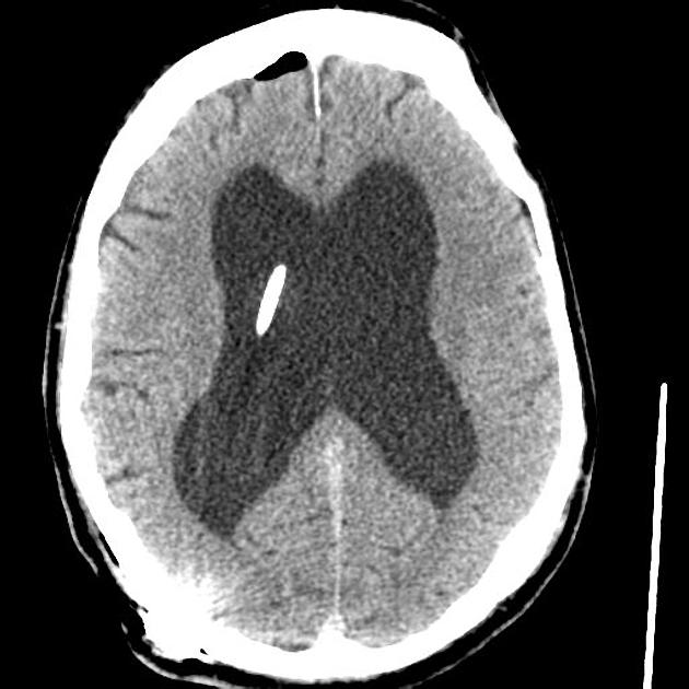
Right posterior approach EVD in situ, with the tip within the body of the right lateral ventricle. Dilated bilateral lateral, third and fourth ventricles is not significantly changed in size. Small amount of bifrontal pneumocephalus is in keeping with post operative status. No intracranial hemorrhage. Previous left frontal craniectomy. Region of encephalomalacia underlying the craniectomy is unchanged.


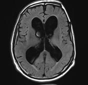

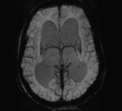
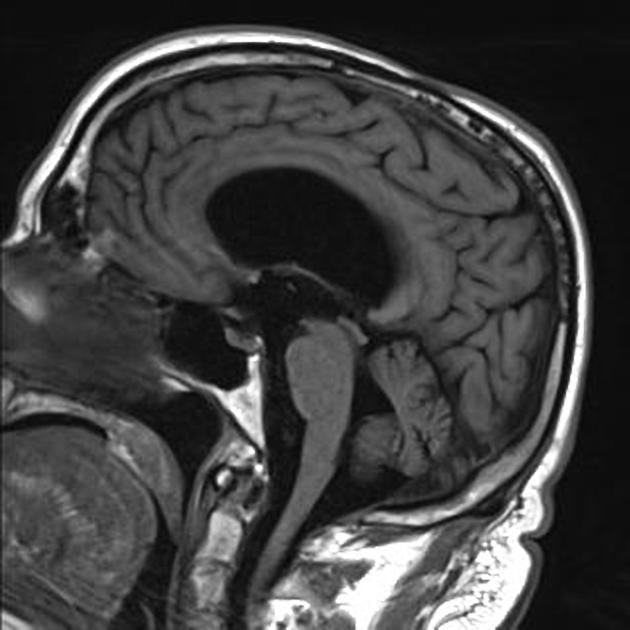
Outside imaging. No report available. Appearances are in compatible with normal pressure hydrocephalous. Clinical picture matched.
Case Discussion
Normal pressure hydrocephalous is one indication where programmable ventriculoperitoneal shunt valves are of particular value. The control radiograph, magnified, will show the settings of the valve which can be correlated with the manufacturer's diagrams.


 Unable to process the form. Check for errors and try again.