
Presentation
Left lower limb deformity.
Patient Data
Age: 6 years
Gender: Female
From the case:
Proximal femoral focal deficiency


Download
Info
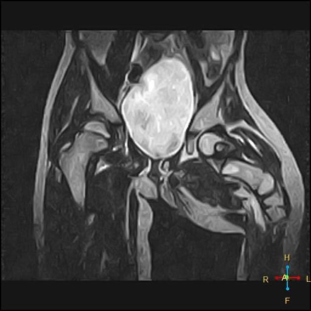
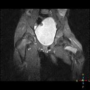
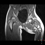
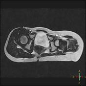
Abnormally short left femur with diaphyseal broadening associated with multiple levels segmentation and pseudoarthrosis.
The femoral head, as well as the left acetabulum, are intact.
No evidence of connection between the femoral head and shaft.
Note the atrophy of the muscles on the left side.
Case Discussion
6 years old female patient with congenitally short left lower limb. MRI of the pelvis revealed abnormal shortening of the left femur with multiple levels segmentation. The femoral head, as well as acetabulum, are intact. Findings are consistent with proximal focal femoral deficiency (PFFD) type A.


 Unable to process the form. Check for errors and try again.