
Presentation
Shortening of right lower limb. The findings on ultrasound raised the concern for right femur aplasia.
Patient Data
Age: 10 months
Gender: Male
From the case:
Proximal femoral focal deficiency - type C

Download
Info
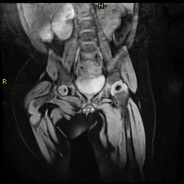
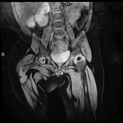
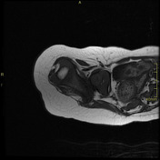
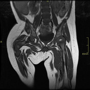
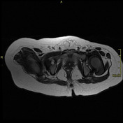
The right femoroacetabular joint is affected with severe coxa vara, identifying:
- Acetabulum present, smaller and slightly dysplastic, with slightly reduced acetabular fossa in size and presence of intraarticular pulvinar fat. The anterior and posterior acetabulum are present with a more rounded and dysplastic morphology. Labrum hypoplasia.
- Presence of epiphyseal proximal femoral ossification nucleus, slightly subluxed superiorly. It is hypoplastic in comparison with the contralateral. It does not present continuity with the femoral shaft.
- The remaining distal femur is shorter with proximal tapering, which ends approximately posterior to the acetabulum in the gluteal fossa.
- There is generalized atrophy of the right thigh muscles.
The distal femur and the right knee joint are congruent with no signs of dysplasia. Right patella hypoplasia. The left femoroacetabular joint is normal in appearance.
From the case:
Proximal femoral focal deficiency - type C
Download
Info
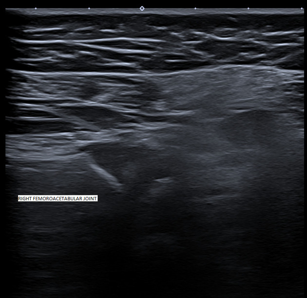
The ultrasound on the right femoracetabular joint shows aplasia / severe hypoplasia of the right femoral head.
Case Discussion
The findings are suggestive of proximal focal femoral deficiency, type C (Atkien classification).
This case is submitted in collaboration with Dr. Emili Inarejos and Dr. Adrià Roset.


 Unable to process the form. Check for errors and try again.