
Presentation
Patient complains of a right breast lump.
Patient Data
Age: 45 years
Gender: Female


Download
Info
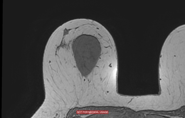
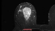
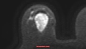
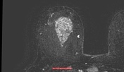
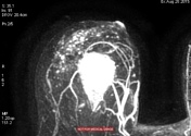
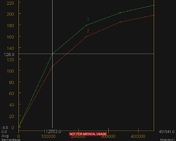
Pear-like shaped mass between upper quadrants (56 mm), with sharp margins. The mass is hyperintense in T2 acquisitions, hypointense in T1 acquisitions. Enhancement is slightly inhomogeneous with an inner core more vividly enhancing. The lesion enhancement shows a type I curve (progressive enhancement). The lesion is hyperintense in DWI acquisitions.
Findings are quite suggestive of a benign lesion.
Case Discussion
Patient underwent a core biopsy which resulted in pseudoangiomatous stromal hyperplasia.


 Unable to process the form. Check for errors and try again.