Breast MRI enhancement curves
Citation, DOI, disclosures and article data
Citation:
Weerakkody Y, Murphy A, Ashraf A, et al. Breast MRI enhancement curves. Reference article, Radiopaedia.org (Accessed on 07 Jan 2025) https://doi.org/10.53347/rID-13886
Permalink:
rID:
13886
Article created:
Disclosures:
At the time the article was created Yuranga Weerakkody had no recorded disclosures.
View Yuranga Weerakkody's current disclosures
Last revised:
Disclosures:
At the time the article was last revised Andrew Murphy had no financial relationships to ineligible companies to disclose.
View Andrew Murphy's current disclosures
Revisions:
12 times, by
10 contributors -
see full revision history and disclosures
Systems:
Sections:
Tags:
Synonyms:
- Breast MRI enhancement kinetics
- MRI enhancement pattern on breast lesions
- Kuhl enhancement curves
- Time intensity curves on breast MRI
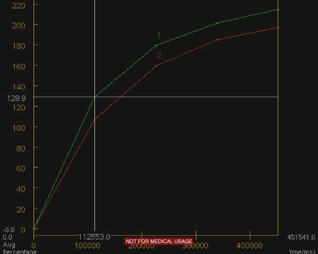
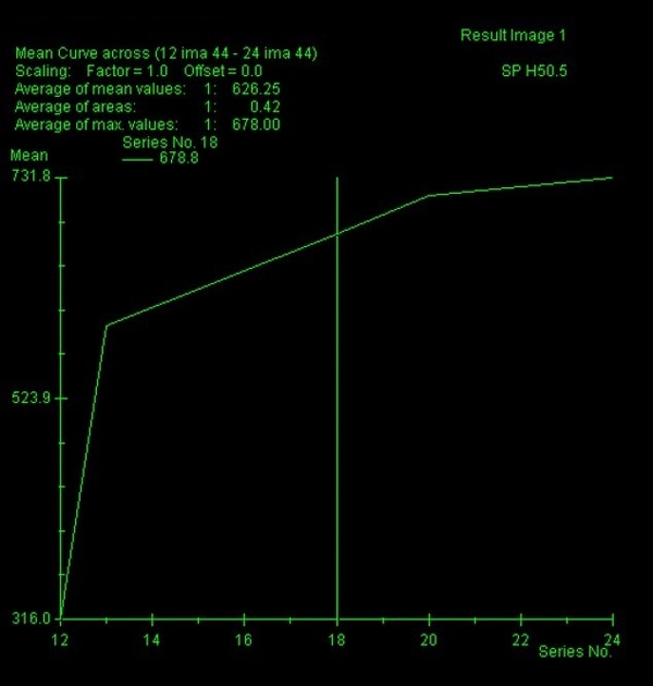
Following administration of gadolinium, there can be three possible enhancement (time intensity) kinetic curves for a lesion on breast MRI (these are also applied in other organs such as prostate MRI). These are sometimes termed the Kuhl enhancement curves.
-
type I curve: progressive or persistent enhancement pattern
- typically shows a continuous increase in signal intensity throughout time
- usually considered benign with only a small proportion of (~9%) of malignant lesions having this pattern
-
type II curve: plateau pattern
- initial uptake followed by the plateau phase towards the latter part of the study
- considered concerning for malignancy
-
type III curve: washout pattern
- has a relatively rapid uptake shows reduction in enhancement towards the latter part of the study
- considered strongly suggestive of malignancy
References
- 1. Moon M, Cornfeld D, Weinreb J. Dynamic contrast-enhanced breast MR imaging. Magn Reson Imaging Clin N Am. 2009;17 (2): 351-62. doi:10.1016/j.mric.2009.01.010 - Pubmed citation
- 2. Macura KJ, Ouwerkerk R, Jacobs MA et-al. Patterns of enhancement on breast MR images: interpretation and imaging pitfalls. Radiographics. 26 (6): 1719-34. doi:10.1148/rg.266065025 - Pubmed citation
- 3. Kuhl C. The current status of breast MR imaging. Part I. Choice of technique, image interpretation, diagnostic accuracy, and transfer to clinical practice. Radiology. 2007;244 (2): 356-78. doi:10.1148/radiol.2442051620 - Pubmed citation
- 4. Kuhl CK. Current status of breast MR imaging. Part 2. Clinical applications. Radiology. 2007;244 (3): 672-91. doi:10.1148/radiol.2443051661 - Pubmed citation
Incoming Links
Articles:
Related articles: Breast imaging
- breast imaging[+][+]
-
mammography
- breast screening
- breast imaging and the technologist
- forbidden (check) areas in mammography
-
mammography views
- craniocaudal view
- mediolateral oblique view
- additional (supplementary) views
- true lateral view
- lateromedial oblique view
- late mediolateral view
- step oblique views
- spot view
- double spot compression view
- magnification view
- exaggerated craniocaudal (axillary) view
- cleavage view
- tangential views
- caudocranial view
- bullseye CC view
- rolled CC view
- elevated craniocaudal projection
- caudal cranial projection
- 20° oblique projection
- inferomedial superolateral oblique projection
- Eklund technique
- normal breast imaging examples
-
mammography
- digital breast tomosynthesis
- breast ultrasound[+][+]
- breast ductography
-
breast MRI
- breast MRI classification flowchart
- breast MRI enhancement curves
- breast morphology[+][+]
- breast intervention[+][+]


 Unable to process the form. Check for errors and try again.