
Presentation
Abdominal pain, fever, jaundice and vomiting.
Patient Data
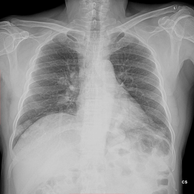
Well defined lobulated radio opacities in the lower zone of the right lung and retrocardiac region of left lower zone.
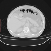

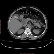
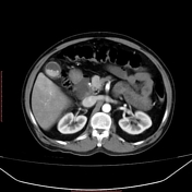
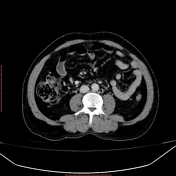
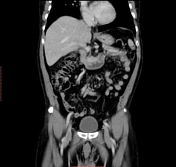
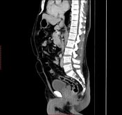

The gall bladder is distended with multiple calculi largest measuring ~ 1.0 x 1.0 cm. Wall thickening (measuring ~ 6.4 mm) showing post contrast enhancement with adjacent pericholecystic fat stranding and thin rim of fluid- features suggestive of cholelithiasis with acute cholecystitis.
Incidentally dilated and tortuous pulmonary vessels in both lower lobes lung parenchyma, the largest in the posterobasal segment of left lower lobe showing avid enhancement.
Case Discussion
Pulmonary arteriovenous malformations are rare anomalies with most patients being asymptomatic.
PAVM can cause dyspnea due to right to left shunt.
All PAVM have an afferent supply usually from the pulmonary artery.
Once diagnosed patient should be treated. Therapeutic options include angiographic embolization with metal coil/balloon occlusion.
PAVM enlarge over a period of time and the morbidity is up to 50% if left untreated.


 Unable to process the form. Check for errors and try again.