
Presentation
Life long history of heavy smoking. Complains of hemoptysis.
Patient Data
Age: 60 years old
Gender: Female
From the case:
Pulmonary coin lesion
Download
Info
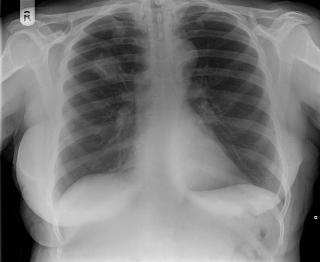
Well defined mass in the right upper lobe.
No mediastinal lymphadenopathy.
Left lung clear.
Case Discussion
A coin lesion on chest x-ray ( this may be a nodule if less than 3cm or in this case a mass as greater than 3cm) requires further assessment.
The differential wide, although the commonest and most important to diagnosis is a bronchial carcinoma.
In the first instance review of previous imaging is indicated. If the lesion was not previously identified then urgent referral to a respiratory physcian is indicated, during this process CT with be performed.


 Unable to process the form. Check for errors and try again.