
Presentation
Cardiac MRI for pulmonary hypertension to rule out cardiac defects or shunts.
Patient Data
Age: 20 years
Gender: Male
Download
Info
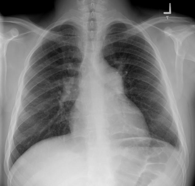
Enlarged pulmonary trunk, lungs and pleural spaces are clear.


Download
Info
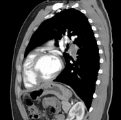
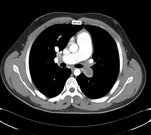
Bilateral segmental pulmonary artery emboli.


Download
Info
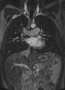
Bilateral pulmonary artery emboli.
Signs of right heart strain are noted:
straightening of the interventricular septum
dilated pulmonary artery, in keeping with the development of pulmonary hypertension
contrast medium reflux into dilated IVC and hepatic veins
Download
Info
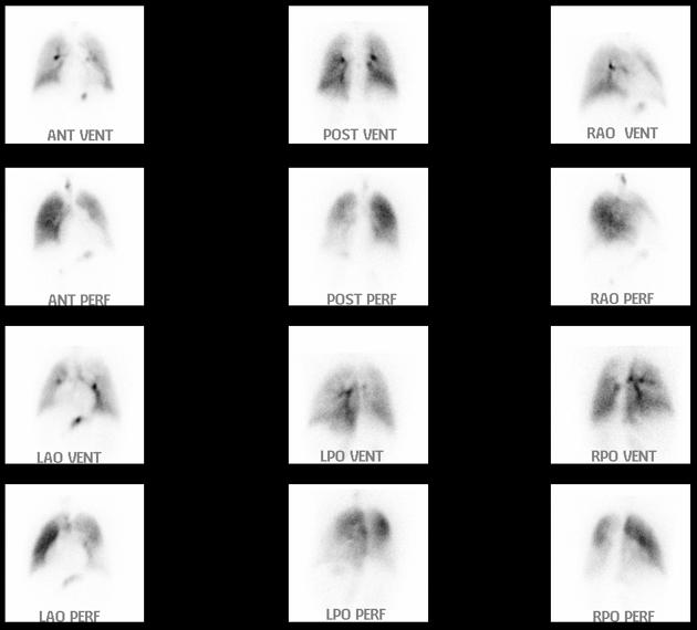
Bilateral major mismatched defects consistent with high probability pulmonary embolism.
Case Discussion
The patient was started on thrombolytic therapy and followed up by CTPA which demonstrated persistent bilateral pulmonary emboli without significant interval changes.


 Unable to process the form. Check for errors and try again.