
Presentation
Recent deep vein thrombosis, dyspnea
Patient Data
Age: 40 years
Gender: Female
From the case:
Pulmonary embolism


Download
Info

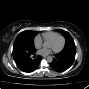
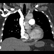
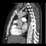
Computed tomography angiography demonstrated multiple filling defects in both pulmonary artery at lobar and segmental level. No signs of pulmonary hypertension or right heart chambers strain.
Mild changes in attenuation in the lung parenchyma.
Incidental right retro-esophageal subclavian artery.
Case Discussion
Findings compatible with acute pulmonary embolism.


 Unable to process the form. Check for errors and try again.