
Presentation
Sudden onset of shortness of breath followed by cough, expectoration and fever.
Patient Data


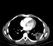

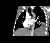
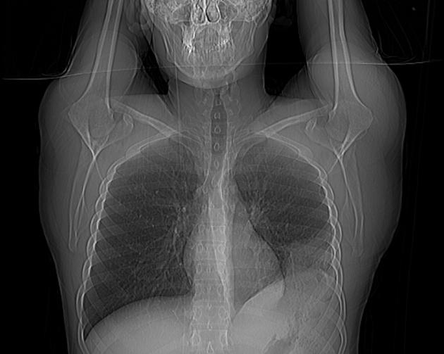
A wedge shape pulmonary consolidation with central cavitation is seen involving the lateral basal segment of left lower lobe. On CT scout image there is classical hampton hump which is peripheral wedge shape area of consolidation. On CT pulmonary angiography there is a filling defect of the segmental pulmonary artery supplying this area.
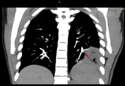
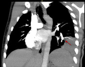

Red arrows illustrate the defect of segmental artery occlusion which supplies the infarcted lateral basal segment of left lower lobe.
Case Discussion
On examination, the patient had tender calf muscle and Doppler study revealed DVT. Pulmonary embolism is a common complication with DVT and can result in pulmonary infarction. Segmental artery thrombosis are difficult to find out and require proper contrast injection and timing.


 Unable to process the form. Check for errors and try again.